. 2018 Jul 17:10:210.
doi: 10.3389/fnagi.2018.00210. eCollection 2018.

Pericyte Structural Remodeling in Cerebrovascular Health and Homeostasis

Affiliations

Pericyte Structural Remodeling in Cerebrovascular Health and Homeostasis

Andrée-Anne Berthiaume et al. Front Aging Neurosci. .

Abstract

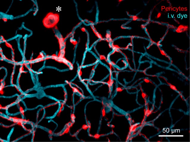
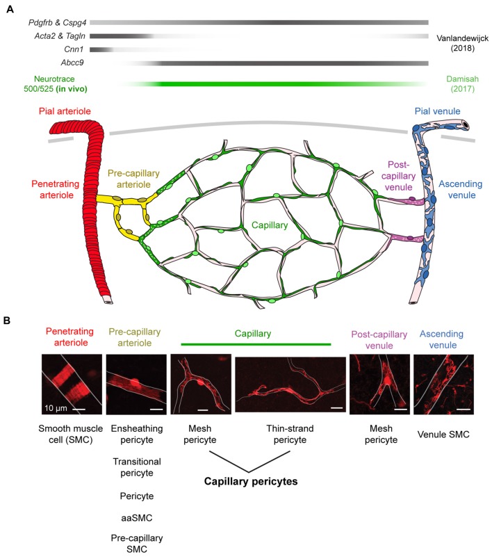
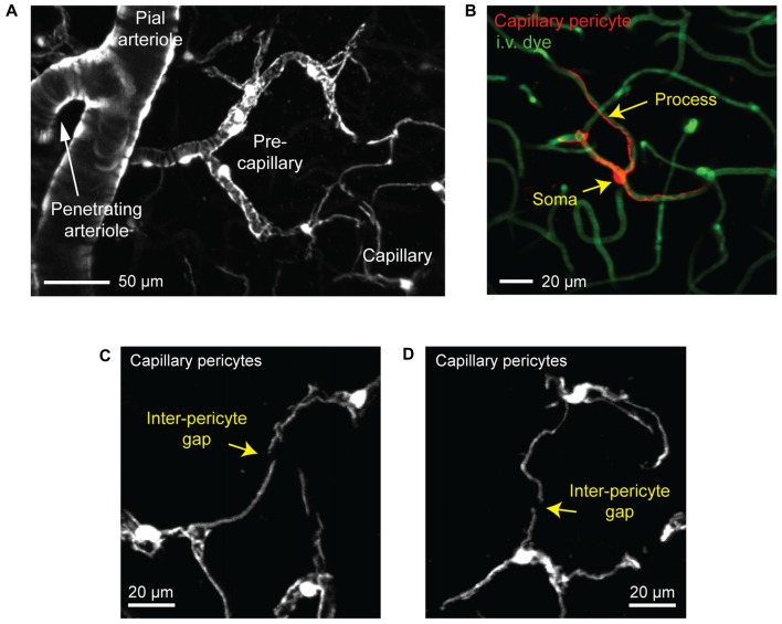
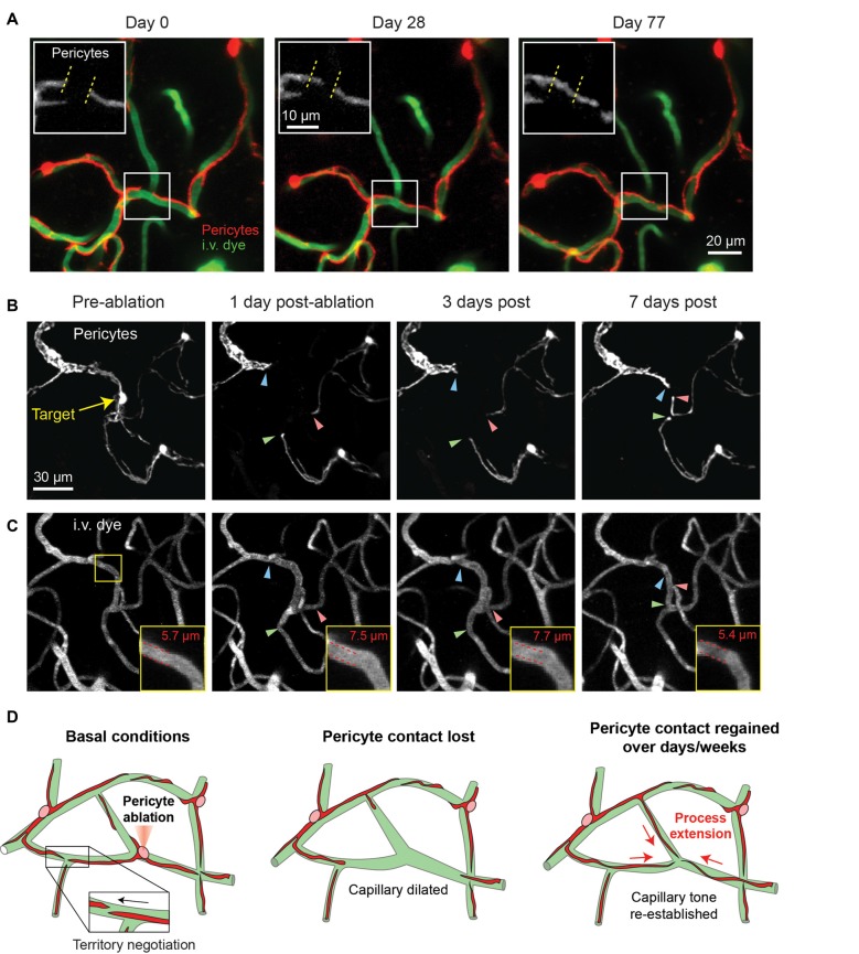
The biology of brain microvascular pericytes is an active area of research and discovery, as their interaction with the endothelium is critical for multiple aspects of cerebrovascular function. There is growing evidence that pericyte loss or dysfunction is involved in the pathogenesis of Alzheimer's disease, vascular dementia, ischemic stroke and brain injury. However, strategies to mitigate or compensate for this loss remain limited. In this review, we highlight a novel finding that pericytes in the adult brain are structurally dynamic in vivo, and actively compensate for loss of endothelial coverage by extending their far-reaching processes to maintain contact with regions of exposed endothelium. Structural remodeling of pericytes may present an opportunity to foster pericyte-endothelial communication in the adult brain and should be explored as a potential means to counteract pericyte loss in dementia and cerebrovascular disease. We discuss the pathophysiological consequences of pericyte loss on capillary function, and the biochemical pathways that may control pericyte remodeling. We also offer guidance for observing pericytes in vivo, such that pericyte structural remodeling can be more broadly studied in mouse models of cerebrovascular disease.

Keywords: Alzheimer’s disease; blood-brain barrier; capillary blood flow; mural cell; neurovascular coupling; pericyte; stroke; two-photon imaging.


Figures

Figure 1
Figure 1
In vivo two-photon microscopy of adult mouse cerebrovasculature. High-resolution image of the capillary network surrounding a penetrating arteriole (*) in the mouse cortex. Mural cells are labeled red through expression of tdTomato driven under the control of the Myosin heavy chain 11 (Myh11) promoter. The vasculature is labeled with intravenously administered 2-MDa FITC-dextran, and pseudo-colored blue. Many inducible and constitutively active Cre drivers are suitable for imaging brain mural cells. For more information, see Hartmann et al. (2015a,b).
Figure 2
Figure 2
Mural cell heterogeneity in the adult mouse cerebrovascular network. (A) (Top) Mural cell gene expression in different zones of the microvasculature in cerebral cortex. Adapted from Vanlandewijck et al. (2018). Neurotrace 500/525 is specifically taken up by capillary pericytes, providing a cell-specific fluorescent label for in vivo imaging (Damisah et al., 2017). (Bottom) Schematic depicting mural cells in seven microvascular zones from pial arterioles to pial venule of cerebral cortex. (B) (Top) Representative images showing morphology of mural cells in five zones below the cortical surface. All scale bars are 10 μm, except the penetrating venule image, which is 50 μm. Adapted from Hartmann et al. (2015a) and Grant et al. (2017). (Bottom) The evolving nomenclature for each cell type. Note that naming of mural cells in the pre-capillary arteriole zone is not universally agreed upon and has become a source of controversy.
Figure 3
Figure 3
Visualizing capillary pericytes with in vivo two-photon imaging. (A) An in vivo image showing shift in mural cell appearance as a pial arteriole branches into an underlying penetrating arteriole, which then ramifies into the pre-capillary arteriole and capillary bed. The image was captured in the cortex of a Myh11-tdTomato mouse and is a maximal projection over 150 μm of cortical thickness. (B) In vivo image of an isolated capillary pericyte following intravenous injection of 2 MDa FITC-dextran to co-label the microvasculature. Image was captured from a NG2-tdTomato mouse. (C,D) Examples of the non-overlapping territories of adjacent capillary pericytes from a Myh11-tdTomato mouse. The location of gaps between two pericyte territories are denoted with arrows.
Figure 4
Figure 4
Pericyte structural remodeling captured with chronic in vivo 2-photon imaging. (A) An example of the structural dynamics of adjacent pericytes under basal conditions. Inset shows the extension of a pericyte process beyond its territory at Day 0, and the corresponding retraction of a neighboring pericyte process. Images are from a Myh11-tdTomato mouse. Adapted from Berthiaume et al. (2018). (B) Two-photon laser ablation of a single pericyte results in the robust extension of immediately adjacent pericyte processes into the vacated territory over 7 days. The image shows the extension of two thin-strand pericytes (green and red arrowheads) and one mesh pericyte (blue arrowhead). Images are from a Myh11-tdTomato mouse. (C) Images of the vasculature, labeled by 2 MDa FITC-dextran, at the site of pericyte ablation (same region as panel B). Inset shows an increased capillary diameter in the vessel segment lacking pericyte coverage, which returns to baseline diameter once pericyte contact is regained suggesting vascular tone. (D) Schematic of pericyte structural remodeling under basal conditions and following acute pericyte ablation.

References

    1. Alarcon-Martinez L., Yilmaz-Ozcan S., Yemisci M., Schallek J., Kılıç K., Can A., et al. . (2018). Capillary pericytes express α-smooth muscle actin, which requires prevention of filamentous-actin depolymerization for detection. Elife 7:e34861. 10.7554/eLife.34861 - DOI - PMC - PubMed
    1. Ando K., Fukuhara S., Izumi N., Nakajima H., Fukui H., Kelsh R. N., et al. . (2016). Clarification of mural cell coverage of vascular endothelial cells by live imaging of zebrafish. Development 143, 1328–1339. 10.1242/dev.132654 - DOI - PMC - PubMed
    1. Arango-Lievano M., Boussadia B., De Terdonck L. D. T., Gault C., Fontanaud P., Lafont C., et al. . (2018). Topographic reorganization of cerebrovascular mural cells under seizure conditions. Cell Rep. 23, 1045–1059. 10.1016/j.celrep.2018.03.110 - DOI - PubMed
    1. Arimura K., Ago T., Kamouchi M., Nakamura K., Ishitsuka K., Kuroda J., et al. . (2012). PDGF receptor β signaling in pericytes following ischemic brain injury. Curr. Neurovasc. Res. 9, 1–9. 10.2174/156720212799297100 - DOI - PubMed
    1. Armulik A., Abramsson A., Betsholtz C. (2005). Endothelial/pericyte interactions. Circ. Res. 97, 512–523. 10.1161/01.res.0000182903.16652.d7 - DOI - PubMed

LinkOut - more resources