. 2018 Jul 17:9:1354.
doi: 10.3389/fmicb.2018.01354. eCollection 2018.

Quorum-Quenching Bacteria Isolated From Red Sea Sediments Reduce Biofilm Formation by Pseudomonas aeruginosa

Affiliations

Quorum-Quenching Bacteria Isolated From Red Sea Sediments Reduce Biofilm Formation by Pseudomonas aeruginosa

Zahid Ur Rehman et al. Front Microbiol. .

Abstract

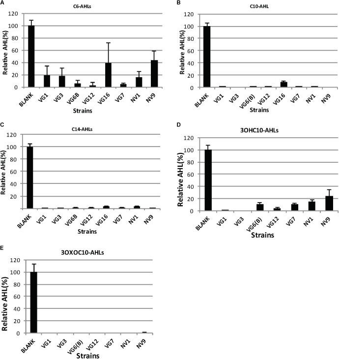
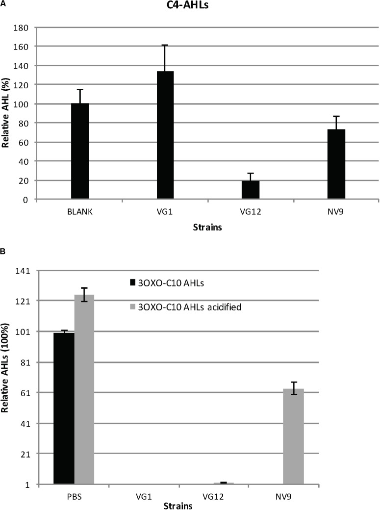
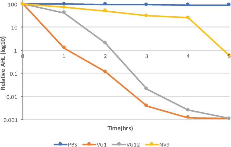
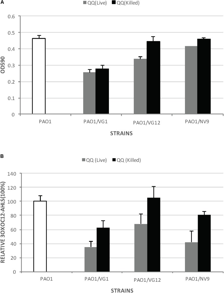
Quorum sensing (QS) is the process by which bacteria communicate with each other through small signaling molecules such as N-acylhomoserine lactones (AHLs). Certain bacteria can degrade AHL molecules by a process called quorum quenching (QQ); therefore, QQ can be used to control bacterial infections and biofilm formation. In this study, we aimed to identify new species of bacteria with QQ activity. Red Sea sediments were collected either from the close vicinity of seagrass or from areas with no vegetation. We isolated 72 bacterial strains, which were tested for their ability to degrade/inactivate AHL molecules. Chromobacterium violaceum CV026-based bioassay was used for the initial screening of isolates with QQ activity. QQ activity was further quantified using high-performance liquid chromatography-tandem mass spectrometry. We found that these isolates could degrade AHL molecules of different acyl chain lengths as well as modifications. 16S-rRNA sequencing of positive QQ isolates showed that they belonged to three different genera. Specifically, two isolates belonged to the genus Erythrobacter; four, Labrenzia; and one, Bacterioplanes. The genome of one representative isolate from each genus was sequenced, and potential QQ enzymes, namely, lactonases and acylases, were identified. The ability of these isolates to degrade the 3OXOC12-AHLs produced by Pseudomonas aeruginosa PAO1 and hence inhibit biofilm formation was investigated. Our results showed that the isolate VG12 (genus Labrenzia) is better than other isolates at controlling biofilm formation by PAO1 and degradation of different AHL molecules. Time-course experiments to study AHL degradation showed that VG1 (genus Erythrobacter) could degrade AHLs faster than other isolates. Thus, QQ bacteria or enzymes can be used in combination with an antibacterial to overcome antibiotic resistance.

Keywords: N-acylhomoserine lactone degradation; Red Sea sediments; biofilm inhibition; marine bacteria; quorum quenching.


Figures

FIGURE 1
FIGURE 1
Degradation of AHLs by the isolated bacteria. The amount of AHLs degraded by different isolates is listed, relative to the negative control. Quantification of AHLs was performed as described in Section “Materials and Methods.” Briefly, AHLs were extracted with ethyl acetate, which was evaporated under a flux of nitrogen gas. The extracted AHLs were re-suspended in acetonitrile and quantified by HPLC-MS. Cell-free PBS was used as the negative control (100%). Values are the mean of three replicates; error bars represent standard deviation. Charts (A–E) illustrates the degradation of C6, C10, C14, 3OXOC10-AHLs, and 3OHC10-AHLs, respectively.
FIGURE 2
FIGURE 2
Degradation and acidification of AHLs. (A) Relative amount of C4-AHLs degraded by the three isolates is given. For quantification, the C4-AHLs were extracted with ethyl acetate and subsequently dried and re-suspended in acetonitrile for injection in HPLC-MS. Cell-free PBS served as the negative control (100%). Experiments were performed in triplicate; error bars represent the standard deviation of the mean value. Student’s t-test showed significant reduction in the amount of C4-AHLs by VG12 (p-value = 0.003) and NV9 (p-value = 0.03). No significant degradation of C4-AHLs by VG1 was observed (p-value = 0.11). (B) Acidification of 3OXOC10-AHLs after incubation with QQ bacteria. Relative amount of AHLs before and after acidification is given. Black bars represent the amount of AHLs after incubation with PBS (negative control is 100%) or QQ bacteria. Gray bars represent the amount of AHLs recovered after acidification. Error bars represent the standard deviation for the three independent replicates.
FIGURE 3
FIGURE 3
Time-course experiment to study AHL degradation. Log10 of the relative amount of AHLs quantified at each time point is given along the y-axis. AHLs were quantified using HPLC–MS, as described in Section “Materials and Methods.” The amount of AHLs at 0 h is considered 100%. Control (PBS) sample and different strains are represented by different colors, as indicated in the legend. Standard deviation at each time point was <10%.
FIGURE 4
FIGURE 4
Biofilm formation by Pseudomonas aeruginosa PAO1 incubated with live and dead QQ strains. (A) This experiment was performed in microtiter plates with membrane inserts for wells, as described in Section “Materials and Methods.” The y-axis indicates the OD590 of the crystal violet bound to the wells. White bars represent biofilm formation by PAO1 without any live or dead QQ bacteria. Gray bars represent biofilm formation by PAO1 incubated with live QQ cells, while black bars represent biofilm formation by PAO1 incubated with dead QQ bacteria. LB broth was used as the negative control. Error bars represent the standard deviation for the three replicates. Student’s t-test was applied to determine significance; p-values: VG1 (0.29), VG12 (0.04), and NV9 (0.09). (B) Relative amount of 3OXOC12-AHLs in the supernatant of PAO1 incubated with live or dead QQ bacteria. The amount of AHLs in the supernatant of PAO1 incubated with live QQ bacteria is shown as gray bars, while that detected in the presence of dead QQ bacteria is shown as black bars. The amount of AHLs produced by PAO1 (without live/dead QQ bacteria) is shown by white bars (100%). Error bars represent the standard deviation. Student’s t-test showed no significant difference in the amount of 3OXOC12-AHLs in the PAO1 supernatant incubated with live/dead VG1 (p-value = 0.16), VG12 (p-value = 0.219), and NV9 (p-value = 0.22).

References

    1. Alam I., Antunes A., Kamau A. A., Ba Alawi W., Kalkatawi M., Stingl U., et al. (2013). INDIGO - INtegrated data warehouse of microbial genomes with examples from the red sea extremophiles. PLoS One 8:e82210. 10.1371/journal.pone.0082210 - DOI - PMC - PubMed
    1. Altschul S. F., Madden T. L., Schaffer A. A., Zhang J., Zhang Z., Miller W., et al. (1997). Gapped BLAST and PSI-BLAST: a new generation of protein database search programs. Nucleic Acids Res. 25 3389–3402. 10.1093/nar/25.17.3389 - DOI - PMC - PubMed
    1. Biebl H., Pukall R., Lunsdorf H., Schulz S., Allgaier M., Tindall B. J., et al. (2007). Description of Labrenzia alexandrii gen. nov., sp. nov., a novel alphaproteobacterium containing bacteriochlorophyll a, and a proposal for reclassification of Stappia aggregata as Labrenzia aggregata comb. nov., of Stappia marina as Labrenzia marina comb. nov. and of Stappia alba as Labrenzia alba comb. nov., and emended descriptions of the genera Pannonibacter, Stappia and Roseibium, and of the species Roseibium denhamense and Roseibium hamelinense. Int. J. Syst. Evol. Microbiol. 571095–1107. 10.1099/ijs.0.64821-0 - DOI - PubMed
    1. Bzdrenga J., Daude D., Remy B., Jacquet P., Plener L., Elias M., et al. (2017). Biotechnological applications of quorum quenching enzymes. Chem. Biol. Interact. 267 104–115. 10.1016/j.cbi.2016.05.028 - DOI - PubMed
    1. Camacho M., Redondo-Gomez S., Rodriguez-Llorente I., Rohde M., Sproer C., Schumann P., et al. (2016). Labrenzia salina sp. nov., isolated from the rhizosphere of the halophyte Arthrocnemum macrostachyum. Int. J. Syst. Evol. Microbiol. 66 5173–5180. 10.1099/ijsem.0.001492 - DOI - PubMed

LinkOut - more resources