

Imaging infective endocarditis: Adherence to a diagnostic flowchart and direct comparison of imaging techniques
- PMID: 30066279
- PMCID: PMC7174257
- DOI: 10.1007/s12350-018-1383-8
Imaging infective endocarditis: Adherence to a diagnostic flowchart and direct comparison of imaging techniques
Abstract
Background: Multimodality imaging is recommended to diagnose infective endocarditis. Value of additional imaging to echocardiography in patients selected by a previously proposed flowchart has not been evaluated.
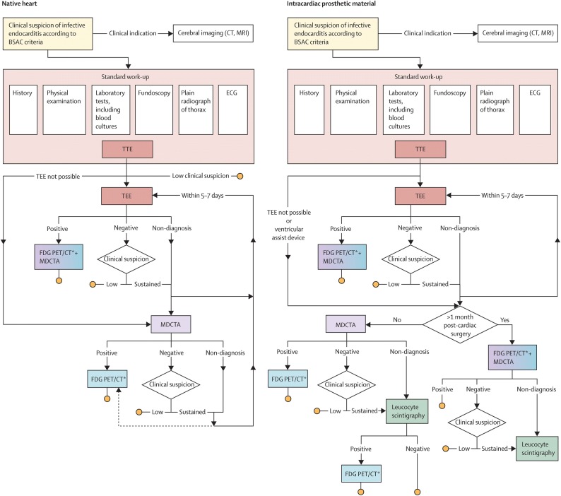
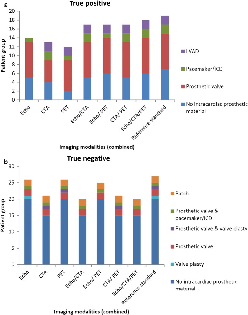
Methods: An observational single-center study was performed. Adult patients suspected of endocarditis/device infection were prospectively and consecutively enrolled from March 2016 to August 2017. Adherence to a diagnostic imaging-in-endocarditis-flowchart was evaluated in 176 patients. Imaging techniques were compared head-to-head in 46 patients receiving echocardiography (transthoracic plus transesophageal), multi-detector computed tomography angiography (MDCTA), and 18F-fluorodeoxyglucose positron emission tomography (FDG-PET/CT).
Results: 69% of patients (121/176) adhered to the flowchart. Sensitivity of echocardiography, MDCTA, FDG-PET/CT in patients without prosthesis was 71%, 57%, 29% (86% when combined), while specificity was 100%, 75%, 100%, respectively. Sensitivity in patients with prosthesis was 75%, 75%, 83%, respectively (100% when combined), while specificity was 86% for all three modalities. Echocardiography performed best in the assessment of vegetations, morphological valve abnormalities/dehiscence, septum defects, and fistula formation. MDCTA performed best in the assessment of abscesses and ventricular assist device infection. FDG-PET/CT performed best in the assessment of cardiac device infection, extracardiac infectious foci, and alternative diagnoses.
Conclusions: This study demonstrates that the evaluated imaging-in-endocarditis-flowchart is applicable in daily clinical practice. Echocardiography, MDCTA, and FDG-PET/CT provide relevant complementary diagnostic information, particularly in patients with intracardiac prosthetic material.
Keywords: CT; Echo; PET; diagnostic and prognostic application; infection; valvular heart disease.
Figures


Comment in
-
Optimizing the diagnostic workup of infective endocarditis: An urgent need for studies focused on defining the decision-making process.J Nucl Cardiol. 2020 Apr;27(2):609-611. doi: 10.1007/s12350-018-1434-1. Epub 2018 Sep 12. J Nucl Cardiol. 2020. PMID: 30209755 No abstract available.
References
-
- Murdoch DR, Corey GR, Hoen B, Miro JM, Fowler VG, Jr, Bayer AS, et al. Clinical presentation, etiology, and outcome of infective endocarditis in the 21st century: The International Collaboration on Endocarditis-Prospective Cohort Study. Arch Intern Med. 2009;169:463–473. doi: 10.1001/archinternmed.2008.603. - DOI - PMC - PubMed
Publication types
MeSH terms
Substances
LinkOut - more resources
Full Text Sources
Other Literature Sources
Medical
