Review
. 2018 Aug;8(1):10.1128/ecosalplus.ESP-0001-2018.
doi: 10.1128/ecosalplus.ESP-0001-2018.

Function and Biogenesis of Lipopolysaccharides

Affiliations
Review

Function and Biogenesis of Lipopolysaccharides

Blake Bertani et al. EcoSal Plus. 2018 Aug.

Abstract

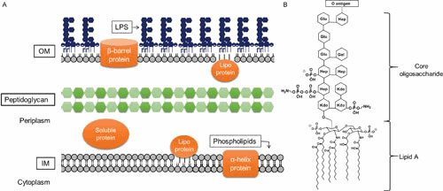
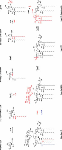
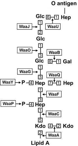
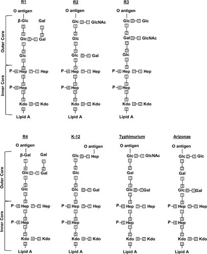
The cell envelope is the first line of defense between a bacterium and the world-at-large. Often, the initial steps that determine the outcome of chemical warfare, bacteriophage infections, and battles with other bacteria or the immune system greatly depend on the structure and composition of the bacterial cell surface. One of the most studied bacterial surface molecules is the glycolipid known as lipopolysaccharide (LPS), which is produced by most Gram-negative bacteria. Much of the initial attention LPS received in the early 1900s was owed to its ability to stimulate the immune system, for which the glycolipid was commonly known as endotoxin. It was later discovered that LPS also creates a permeability barrier at the cell surface and is a main contributor to the innate resistance that Gram-negative bacteria display against many antimicrobials. Not surprisingly, these important properties of LPS have driven a vast and still prolific body of literature for more than a hundred years. LPS research has also led to pioneering studies in bacterial envelope biogenesis and physiology, mostly using Escherichia coli and Salmonella as model systems. In this review, we will focus on the fundamental knowledge we have gained from studies of the complex structure of the LPS molecule and the biochemical pathways for its synthesis, as well as the transport of LPS across the bacterial envelope and its assembly at the cell surface.
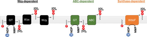
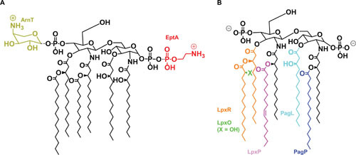
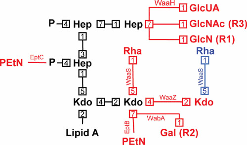
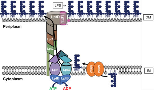

Figures

Figure 1
Figure 1
Architecture of the Gram-negative cell envelope. (A) Depiction of the Gram-negative cell envelope and its components. The inner membrane (IM) contains phospholipids, while the outer membrane (OM) contains phospholipids in the inner leaflet and lipopolysaccharide (LPS) in the outer leaflet. (B) Structure of prototypical LPS produced by E. coli (shown is the core structure associated with core type K-12).
Figure 2
Figure 2
Lipid A biosynthesis pathway. Modifications to the preceding structure made by each enzyme in the pathway are marked in red, with the exception of the last step, where the modifications made by LpxL and LpxM are colored in red and blue, respectively. Donor molecules are not shown. At low temperatures, LpxP acts instead of LpxL to add a C16:1 palmitoleoyl group instead of a lauroyl group.
Figure 3
Figure 3
Structure and biosynthetic enzymes of the E. coli K-12 core oligosaccharide. Numbers represent bond positions between sugars. Note that nonstoichiometric modifications are not shown. All linkages are α-anomeric unless preceded by the β symbol, which specifies the β-anomeric state. Enzyme names are boxed, with arrows indicating the linkages they catalyze. It is worth noting that, while the O-antigen ligation site is indicated, E. coli K-12 does not typically produce O antigen because of an ancestral mutation that inactivates its synthesis.
Figure 4
Figure 4
Structure of various core oligosaccharides. Shown are the known core types in E. coli (R1 to R4, K-12) and S. enterica serovar Typhimurium and S. enterica serovar Arizonae IIIA. Numbers represent bond positions between sugars. Note that nonstoichiometric modifications are not shown. All linkages are α-anomeric unless preceded by the β symbol, which specifies the β-anomeric state.
Figure 5
Figure 5
Summary of different O-antigen synthesis pathways. GT stands for glycosyltransferase, and for the purposes of illustration represents all GTs required to generate the O antigen. [O] represents a repeating unit of the O antigen, while the subscript represents the number of repeats present (n being an arbitrary integer). Individual sugar units are represented by “S” inside a hexagon, and are shown bound to an arbitrary nucleotide carrier NDP. The lipid carrier is Und-P.
Figure 6
Figure 6
Modifications to the structure of lipid A. Shown are modifications made to lipid A described in the text, alongside the enzymes that mediate them in corresponding colors. (A) Modifications made to the glucosamine phosphates. While shown in their preferred positions, it is possible for either phosphate to be modified with either substituent. (B) Modifications made to the acyl groups. The X indicated for LpxO is a hydroxyl (-OH) when LpxO is active, and a hydrogen (-H) when it is not. When acyl chains are removed, the cleaved bond is shown as a dotted line. It is important to note that LpxP is part of the conserved Raetz pathway but only active at low temperatures, at which LpxP substitutes for LpxL to add a C16:1 palmitoleoyl group instead of a lauroyl group.
Figure 7
Figure 7
Modifications to the structure of the core oligosaccharide. Shown is the conserved inner core oligosaccharide (and its linkage to lipid A) in black, with potential modifications being indicated in red (although the alternate rhamnose modification by WaaS when the second Kdo is modified with PEtN by EptB is shown in blue). Numbers represent bond positions between sugars. Enzymes mediating modifications are next to each linkage, and, when not associated with E. coli K-12, core type associations are listed in parentheses beside the modification.
Figure 8
Figure 8
Transport of LPS across the cell envelope. Shown are representations of MsbA, which mediates the transport of core-lipid A across the IM, and the Lpt complex (LptB2FGCADE), which mediates LPS extraction from the IM and its transport through the periplasm and OM. As described in the text, the O antigen can be synthesized on Und-P and transported across the IM by different pathways (Fig. 5). If made, the O antigen is ligated to core-lipid A in the periplasmic leaflet of the IM by WaaL (not shown).

References

    1. Silhavy TJ, Kahne D, Walker S. 2010. The bacterial cell envelope. Cold Spring Harb Perspect Biol 2:a000414. 10.1101/cshperspect.a000414. [PubMed] - DOI - PMC - PubMed
    1. Bos MP, Robert V, Tommassen J. 2007. Biogenesis of the gram-negative bacterial outer membrane. Annu Rev Microbiol 61:191–214. [PubMed] - PubMed
    1. Kamio Y, Nikaido H. 1976. Outer membrane of Salmonella typhimurium: accessibility of phospholipid head groups to phospholipase c and cyanogen bromide activated dextran in the external medium. Biochemistry 15:2561–2570. [PubMed] - PubMed
    1. Zhang G, Meredith TC, Kahne D. 2013. On the essentiality of lipopolysaccharide to Gram-negative bacteria. Curr Opin Microbiol 16:779–785. [PubMed] - PMC - PubMed
    1. Nikaido H. 2003. Molecular basis of bacterial outer membrane permeability revisited. Microbiol Mol Biol Rev 67:593–656. [PubMed] - PMC - PubMed

Publication types

MeSH terms