Review
. 2018 Jul 26;113(9):e180212.
doi: 10.1590/0074-02760180212.

What are the advantages of living in a community? A microbial biofilm perspective!

Affiliations
Review

What are the advantages of living in a community? A microbial biofilm perspective!

André Luis Souza Dos Santos et al. Mem Inst Oswaldo Cruz. .

Abstract

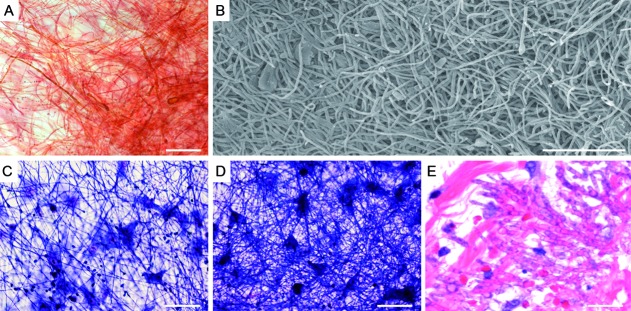
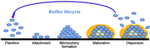
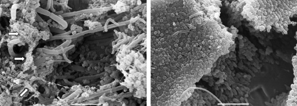
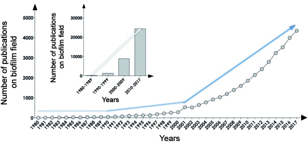
Biofilm formation is the preferred mode of growth lifestyle for many microorganisms, including bacterial and fungal human pathogens. Biofilm is a strong and dynamic structure that confers a broad range of advantages to its members, such as adhesion/cohesion capabilities, mechanical properties, nutritional sources, metabolite exchange platform, cellular communication, protection and resistance to drugs (e.g., antimicrobials, antiseptics, and disinfectants), environmental stresses (e.g., dehydration and ultraviolet light), host immune attacks (e.g., antibodies, complement system, antimicrobial peptides, and phagocytes), and shear forces. Microbial biofilms cause problems in the hospital environment, generating high healthcare costs and prolonged patient stay, which can result in further secondary microbial infections and various health complications. Consequently, both public and private investments must be made to ensure better patient management, as well as to find novel therapeutic strategies to circumvent the resistance and resilience profiles arising from biofilm-associated microbial infections. In this work, we present a general overview of microbial biofilm formation and its relevance within the biomedical context.


Figures

Fig. 1:
Fig. 1:. biofilm formation by the filamentous fungus, Scedosporium apiospermum, an opportunistic human pathogen, on both abiotic and biotic surfaces. The biofilm on the polystyrene substrate was detected by safranin staining (A) and by scanning electron microscopy (B). The co-culture of S. apiospermum conidia with lung epithelial A549 cells for 24 h (C) and 48 h (D) revealed a dense mycelial mass (a typical biofilm structure) covering the lung epithelial cells, as evidenced by Giemsa staining. Histopathological section stained by hematoxylin and eosin showing many hyphae in the skin biopsy from human infected tissue (E). Bars, 50 µm. For more detail see Mello et al. Image credit: Fig. E was kindly gifted by Dr Stacy Beal, Assistant Professor, University of Florida, College of Medicine, Department of Pathology, Immunology and Laboratory Medicine, USA.
Fig. 2:
Fig. 2:. biofilm development steps produced by microorganisms. The image indicates the adherence of a microbial community to the surface, followed by microcolony formation, the maturation of the biofilm 3D architecture (note the presence of different cells, including the persisters, which are represented by light blue circles) with the presence of an extracellular polymeric matrix (represented by an orange cover) and, finally, its dispersion, which can lead to the colonisation and formation of a new biofilm structure in a distinct place, as well as the persistence of the infectious disease.
Fig. 3:
Fig. 3:. polymicrobial biofilms detected on orthodontic appliances visualised by scanning electron microscopy (SEM). The SEM images reveal the presence of different microbial groups, including bacteria and fungi, and an extracellular matrix (arrows). Bars, 5 µm.
Fig. 4:
Fig. 4:. biofilm publications. The search was performed in the PubMed database (www.pubmed.com) on April 1st, 2018, using the keyword “biofilm”, and only papers published in English containing this word in the title and/or in the abstract were selected for analysis. Subsequently, the list of results was exported to the EndNote software (version X1), using the “Output Records” tool in order to eliminate duplicated references by means of the “Find Duplicates” tool. The graphic shows the absolute number of publications in the biofilm field during the period 1980-2017 (blue lines denote the increasing number of publications over the years). Note the substantial increase in the number of publications across the timeline, especially comparing the period of 1980-1989 with subsequent periods.
Fig. 5:
Fig. 5:. publication metrics in the anti-biofilm field. The search was performed in the PubMed database (www.pubmed.com) on April 1st, 2018, using the keywords “anti-biofilm”, “anti-biofilm agents”, and “anti-biofilm strategies”, and only papers published in English containing these words were selected for analysis. The graphic shows the number of publications in these biofilm arenas during the period 2001-2017, which is the most representative period for publications on biofilms (Fig. 4).

References

    1. Stevison LS, Sefick S, Rushton C, Graze RM. Recombination rate plasticity revealing mechanisms by design. Philos Trans R Soc Lond B Biol Sci. 2017;372(1712):20160459–20160459. - PMC - PubMed
    1. Hiltunen T, Virta M, Laine AL. Antibiotic resistance in the wild an eco-evolutionary perspective. Philos Trans R Soc Lond B Biol Sci. 2017;372(1712):20160039–20160039. - PMC - PubMed
    1. Høiby N. A short history of microbial biofilms and biofilm infections. APMIS. 2017;125(4):272–275. - PubMed
    1. Flemming HC, Wingender J. The biofilm matrix. Nat Rev Microbiol. 2010;8(9):623–633. - PubMed
    1. Lopez D, Vlamakis H, Kolter R. Biofilms. Cold Spring Harb Perspect Biol. 2010;2(7):a000398–a000398. - PMC - PubMed