Chromoblastomycosis: an etiological, epidemiological, clinical, diagnostic, and treatment update
- PMID: 30066754
- PMCID: PMC6063100
- DOI: 10.1590/abd1806-4841.20187321
Chromoblastomycosis: an etiological, epidemiological, clinical, diagnostic, and treatment update
Abstract
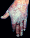
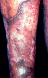
Chromoblastomycosis is a chronic, granulomatous, suppurative mycosis of the skin and subcutaneous tissue caused by traumatic inoculation of dematiaceous fungi of the family Herpotrichiellaceae. The species Fonsecaea pedrosoi and Cladophialophora carrionii are prevalent in regions where the disease is endemic. Chromoblastomycosis lesions are polymorphous: verrucous, nodular, tumoral, plaque-like, and atrophic. It is an occupational disease that predominates in tropical and subtropical regions, but there have been several reports of cases in temperate regions. The disease mainly affects current or former farm workers, mostly males, and often leaving disabling sequelae. This mycosis is still a therapeutic challenge due to frequent recurrence of lesions. Patients with extensive lesions require a combination of pharmacological and physical therapies. The article provides an update of epidemiological, clinical, diagnostic, and therapeutic features.
Conflict of interest statement
Conflict of interest: None.
Figures


References
-
- McGinnis MR. Chromoblastomycosis and phaeohyphomycosis: new concepts, diagnosis, and mycology. J Am Acad Dermatol. 1983;8:1–16. - PubMed
-
- Rubin HA, Bruce S, Rosen T, McBride ME. Evidence for percutaneous inoculation as the mode of transmission for chromoblastomycosis. J Am Acad Dermatol. 1991;25:951–954. - PubMed
-
- Terra F, Torres M, Fonseca Filho O. Novo tipo de dermatite verrucosa; micose por Acrotheca com associado de leishmaniose. Brasil Medico. 1922;36:363–368.
-
- Odds FC, Arai T, Disalvo AF, Evans EG, Hay RJ, Randhawa HS. Nomenclature of fungal diseases: a report and recommendations from a Sub-Committee of the International Society for Human and Animal Mycology (ISHAM) J Med Vet Mycol. 1992;30:1–10. - PubMed
Publication types
MeSH terms
LinkOut - more resources
Full Text Sources
Other Literature Sources

