

Functional neuroanatomy of peripheral inflammatory physiology: A meta-analysis of human neuroimaging studies
- PMID: 30067939
- PMCID: PMC6363360
- DOI: 10.1016/j.neubiorev.2018.07.013
Functional neuroanatomy of peripheral inflammatory physiology: A meta-analysis of human neuroimaging studies
Abstract
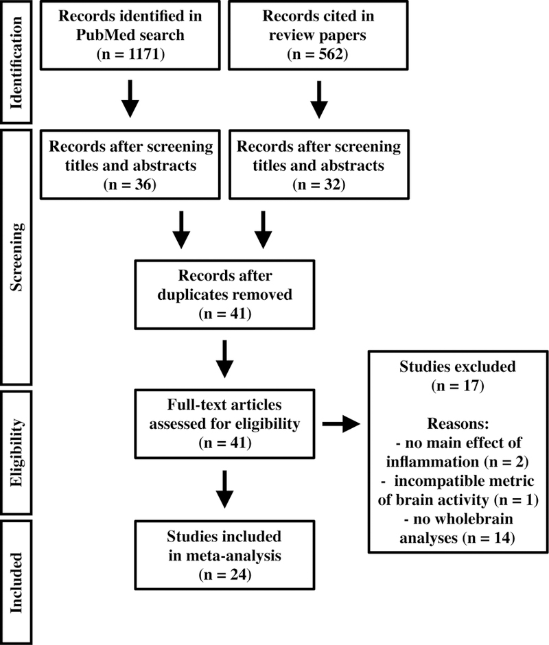
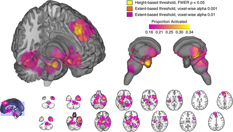
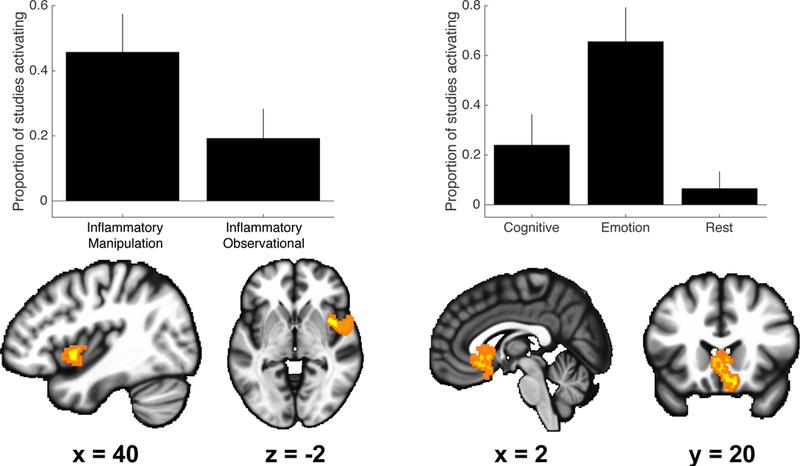
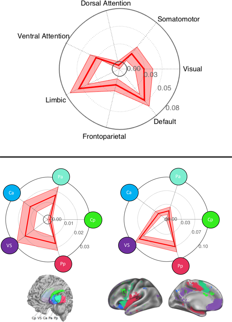
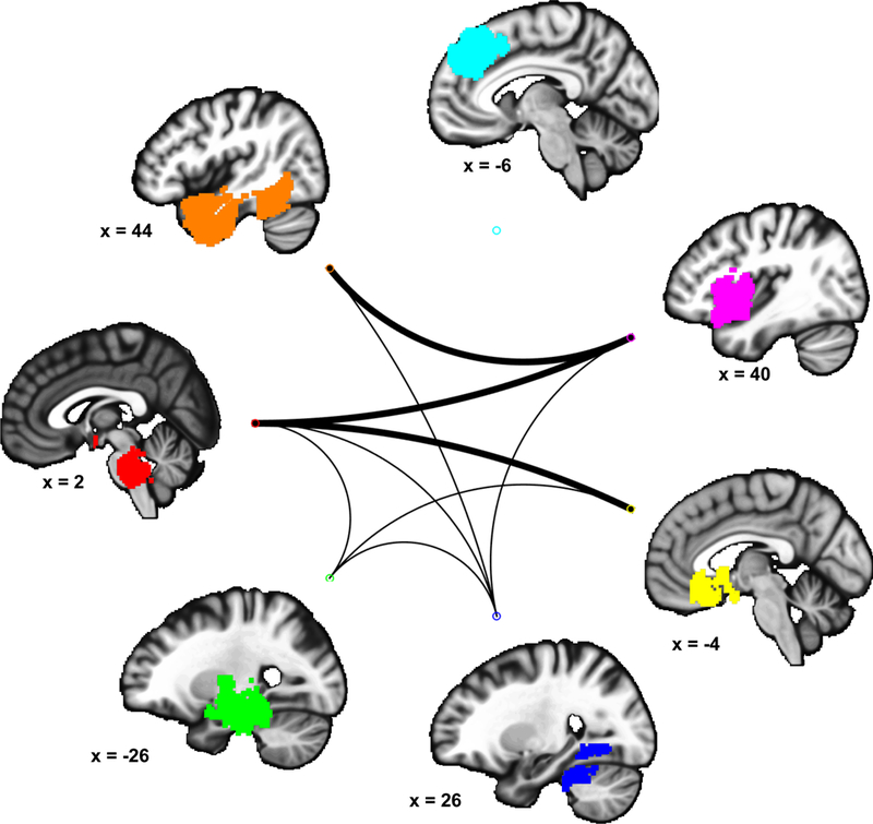
Communication between the brain and peripheral mediators of systemic inflammation is implicated in numerous psychological, behavioral, and physiological processes. Functional neuroimaging studies have identified brain regions that associate with peripheral inflammation in humans, yet there are open questions about the consistency, specificity, and network characteristics of these findings. The present systematic review provides a meta-analysis to address these questions. Multilevel kernel density analysis of 24 studies (37 statistical maps; 264 coordinates; 457 participants) revealed consistent effects in the amygdala, hippocampus, hypothalamus, striatum, insula, midbrain, and brainstem, as well as prefrontal and temporal cortices. Effects in some regions were specific to particular study designs and tasks. Spatial pattern analysis revealed significant overlap of reported effects with limbic, default mode, ventral attention, and corticostriatal networks, and co-activation analyses revealed functional ensembles encompassing the prefrontal cortex, insula, and midbrain/brainstem. Together, these results characterize brain regions and networks associated with peripheral inflammation in humans, and they provide a functional neuroanatomical reference point for future neuroimaging studies on brain-body interactions.
Keywords: Brain; Functional magnetic resonance imaging; Immunity; Inflammation; Meta-analysis; Multilevel kernel density analysis; Neuroimaging; Positron emission tomography; Stress.
Copyright © 2018 Elsevier Ltd. All rights reserved.
Conflict of interest statement
The authors attest that they have no conflicts of interest.
Figures
Similar articles
-
Functional neuroimaging studies of human emotions.CNS Spectr. 2004 Apr;9(4):258-66. doi: 10.1017/s1092852900009196. CNS Spectr. 2004. PMID: 15048050 Review.
-
Neuroimaging procedures and related acquisitions in bipolar disorder: state of the art.Riv Psichiatr. 2014 Jan-Feb;49(1):2-11. doi: 10.1708/1407.15619. Riv Psichiatr. 2014. PMID: 24572578 Review.
-
Systemic inflammation and resting state connectivity of the default mode network.Brain Behav Immun. 2017 May;62:162-170. doi: 10.1016/j.bbi.2017.01.013. Epub 2017 Jan 23. Brain Behav Immun. 2017. PMID: 28126500 Free PMC article.
-
Altered corticostriatal functional connectivity in obsessive-compulsive disorder.Arch Gen Psychiatry. 2009 Nov;66(11):1189-200. doi: 10.1001/archgenpsychiatry.2009.152. Arch Gen Psychiatry. 2009. PMID: 19884607
-
Chronic cigarette smoking is linked with structural alterations in brain regions showing acute nicotinic drug-induced functional modulations.Behav Brain Funct. 2016 Jun 2;12(1):16. doi: 10.1186/s12993-016-0100-5. Behav Brain Funct. 2016. PMID: 27251183 Free PMC article.
Cited by
-
Biological factors influencing depression in later life: role of aging processes and treatment implications.Transl Psychiatry. 2023 May 10;13(1):160. doi: 10.1038/s41398-023-02464-9. Transl Psychiatry. 2023. PMID: 37160884 Free PMC article. Review.
-
Links between brain neuroimaging and blood inflammatory markers in urological chronic pelvic pain syndrome.Physiol Behav. 2023 Nov 1;271:114358. doi: 10.1016/j.physbeh.2023.114358. Epub 2023 Sep 26. Physiol Behav. 2023. PMID: 37769862 Free PMC article.
-
Medial prefrontal cortex connectivity with the nucleus accumbens is related to HIV serostatus, perceptions of psychological stress, and monocyte expression of TNF-a.Brain Behav Immun Health. 2024 Aug 22;41:100844. doi: 10.1016/j.bbih.2024.100844. eCollection 2024 Nov. Brain Behav Immun Health. 2024. PMID: 39328275 Free PMC article.
-
Towards a multidimensional model of inflamed depression.Brain Behav Immun Health. 2022 Nov 20;26:100564. doi: 10.1016/j.bbih.2022.100564. eCollection 2022 Dec. Brain Behav Immun Health. 2022. PMID: 36439056 Free PMC article.
-
Clinically relevant effects of Mindfulness-Based Stress Reduction in individuals with asthma.Brain Behav Immun Health. 2022 Sep 14;25:100509. doi: 10.1016/j.bbih.2022.100509. eCollection 2022 Nov. Brain Behav Immun Health. 2022. PMID: 36177306 Free PMC article.
References
-
- Agresti A, 2002. Inference for contingency tables. Categ. Data Anal Second Ed. 70–114.
-
- Banks WA, Kastin AJ, 1991. Blood to brain transport of interleukin links the immune and central nervous systems. Life Sci 48, PL117–121. - PubMed
-
- Baron RM, Kenny DA, 1986. The moderator–mediator variable distinction in social psychological research: Conceptual, strategic, and statistical considerations. J. Pers. Soc. Psychol 51, 1173. - PubMed
Publication types
MeSH terms
Grants and funding
LinkOut - more resources
Full Text Sources
Other Literature Sources
