Comparative Study
. 2018 Aug 1;19(1):573.
doi: 10.1186/s12864-018-4957-6.

A map of gene expression in neutrophil-like cell lines

Affiliations
Comparative Study

A map of gene expression in neutrophil-like cell lines

Esther Rincón et al. BMC Genomics. .

Abstract

Background: Human neutrophils are central players in innate immunity, a major component of inflammatory responses, and a leading model for cell motility and chemotaxis. However, primary neutrophils are short-lived, limiting their experimental usefulness in the laboratory. Thus, human myeloid cell lines have been characterized for their ability to undergo neutrophil-like differentiation in vitro. The HL-60 cell line and its PLB-985 sub-line are commonly used to model human neutrophil behavior, but how closely gene expression in differentiated cells resembles that of primary neutrophils has remained unclear.

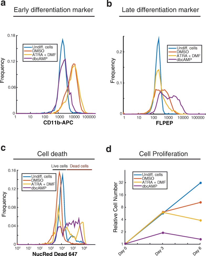
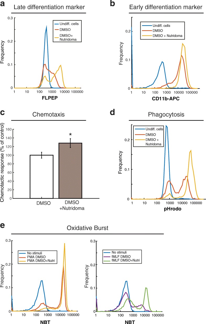
Results: In this study, we compared the effectiveness of differentiation protocols and used RNA sequencing (RNA-seq) to compare the transcriptomes of HL-60 and PLB-985 cells with published data for human and mouse primary neutrophils. Among commonly used differentiation protocols for neutrophil-like cell lines, addition of dimethyl sulfoxide (DMSO) gave the best combination of cell viability and expression of markers for differentiation. However, combining DMSO with the serum-free-supplement Nutridoma resulted in increased chemotactic response, phagocytic activity, oxidative burst and cell surface expression of the neutrophil markers FPR1 and CD11b without a cost in viability. RNA-seq analysis of HL-60 and PLB-985 cells before and after differentiation showed that differentiation broadly increases the similarity in gene expression between the cell lines and primary neutrophils. Furthermore, the gene expression pattern of the differentiated cell lines correlated slightly better with that of human neutrophils than the mouse neutrophil pattern did. Finally, we created a publicly available gene expression database that is searchable by gene name and protein domain content, where users can compare gene expression in HL-60, PLB-985 and primary human and mouse neutrophils.

Conclusions: Our study verifies that a DMSO-based differentiation protocol for HL-60 and PLB-985 cell lines gives superior differentiation and cell viability relative to other common protocols, and indicates that addition of Nutridoma may be preferable for studies of chemotaxis, phagocytosis, or oxidative burst. Our neutrophil gene expression database will be a valuable tool to identify similarities and differences in gene expression between the cell lines and primary neutrophils, to compare expression levels for genes of interest, and to improve the design of tools for genetic perturbations.

Keywords: Chemotaxis; Database; Differentiation protocol; Neutrophil; Neutrophil-like cell line; RNA-seq.


Conflict of interest statement

Ethical approval for the study of neutrophils from healthy adult volunteers was granted by the Institutional Review Board (IRB) from the University of California, Davis (IORG0000251). All participants gave written, informed consent.

Not applicable

The authors declare that they have no competing interests.

Springer Nature remains neutral with regard to jurisdictional claims in published maps and institutional affiliations.

Figures

Fig. 1
Fig. 1
DMSO-based differentiation protocol gives the best differentiation potential based on expression of differentiation markers and cell viability. PLB-985 cells were differentiated into a neutrophil-like state by culturing in different media (DMSO, ATRA + DMF, or dbcAMP) for 6 days. Undifferentiated cells were also analyzed. Cells were stained with an antibody against CD11b, chosen as an early differentiation marker (a), with FLPEP (a fluorescent ligand of FPR1), chosen as a late differentiation marker (b), or with NucRed Dead 647 Probe, to measure cell death (c). Samples were measured by cytometry and data was analyzed using MATLAB. d Cell growth was monitored at days zero, three, and six of the differentiation protocols, using the trypan blue dye exclusion test. The number of cells were normalized to the initial number of cells. These experiments were performed three times and a representative experiment is shown
Fig. 2
Fig. 2
Replacing serum with Nutridoma during differentiation increases FPR1 surface expression and chemotactic efficiency. PLB-985 cells were differentiated into a neutrophil-like state by culturing in media supplemented with 1.3% DMSO and 9% FBS or supplemented with 1.3% DMSO, 2% Nutridoma and 0.5% FBS, for 6 days. Then cells were stained with FLPEP (a) or an antibody against CD11b (b) and measured by cytometry. Data was analyzed using MATLAB. The experiment was repeated three times and a representative experiment is shown. (c) Cells differentiated as in (a) and (b) were plated under agarose and analyzed in an automated chemotaxis assay by time-lapse microscopy with chemoattractant uncaging of Nv-fMLF. A cell directionality parameter measuring the angular bias of cell movement towards the gradient source is shown. This experiment was performed three times. Error bars represent the standard error of the mean. Images and statistics were processed using custom MATLAB software. Asterisk indicates a p-value of less than 0.05 using a t-test. d Cells differentiated as in (a) and (b) were mixed in suspension with pHrodo Green-labeled dead Staphylococcus aureus bioparticles for 2 h at 37 degrees. Phagocytosis of the particles was then analyzed by cytometry. e PLB-985 cells were differentiated into a neutrophil-like state as in (a) and (b). Cells were then incubated with NBT solution and 100 ng/mL of PMA or 1 μM fMLF, at 37 °C for 15 min, and measured by cytometry
Fig. 3
Fig. 3
Comparison of differentiated PLB-985 cells with primary neutrophils. a Undifferentiated PLB-985 cells, PLB-985 cells differentiated with DMSO and Nutridoma, and primary neutrophils were stained with antibody against CD11b and analyzed by cytometry. Staining of primary neutrophils with an isotype control antibody is also shown. b The same cell samples were stained with FLPEP and analyzed by cytometry. Unstained samples are also shown for comparison. c Phagocytosis of pHrodo Green-labeled dead Staphylococcus aureus bioparticles. Negative controls in which no particles were present are also shown. d-f Differentiated PLB-985 cells and primary neutrophils were plated under agarose and analyzed in an automated chemotaxis assay by time-lapse microscopy with chemoattractant uncaging of Nv-fMLF. Mean cell speed was measured both before (d) and after (e) generation of an fMLF gradient. A cell directionality parameter measuring the angular bias of cell movement towards the gradient source is shown (f). An angular bias of 90 degrees indicates perfect directionality, and zero degrees indicates random orientation. This experiment was performed three times. Error bars represent the standard error of the mean. Asterisk indicates a p-value of less than 0.05 and double asterisks indicate a p-value of less than 0.01 using a t-test. g Histograms of the instantaneous directionality of individual cells is shown for the same experiments analyzed in (f). Here an angle of zero indicates optimal directionality, and 180 degrees indicates movement in the opposite direction. A third curve (yellow) indicates simulated data for a population of neutrophils in which only 70% of cells express the receptor FPR1
Fig. 4
Fig. 4
Transcriptional profiles of HL-60 and PLB-985 cells before and after differentiation and comparison to that of primary neutrophils. a Experimental design: HL-60 and PLB-985 cells were differentiated by culturing in media supplemented with 1.3% DMSO and 9% FBS or 1.3% DMSO, 2% Nutridoma and 0.5% FBS. Total RNA of cells harvested at the indicated time points was isolated. PolyA enrichment and RNA-seq were performed by Applied Biological Materials Inc. through their Total RNA Sequencing service. RNA-seq data was subjected to our analysis pipeline as described in Methods. b Heat map of Spearman correlations between transcriptional profiles of undifferentiated and differentiated HL-60 and PLB-985 cells and human and mouse primary neutrophils, organized by unbiased hierarchical clustering. Color bar indicates correlation strength. Each row/column represents an independent sample. For the cell line data, the labels d2, d4, and d6 indicate 2, 4, and 6 days of differentiation, respectively. For the Thomas et al. data, the labels s1, s2, s3, and s4 indicate sample numbers, which correspond to the sample numbers in Table 2
Fig. 5
Fig. 5
PLB-985 cell differentiation results in neutrophil-like gene expression patterns. a Histogram of log10 fold expression changes between replicate measurements of undifferentiated PLB-985 cells (ND), and between undifferentiated cells and those differentiated with either 1.3% DMSO and 9% FBS (DMSO) or DMSO + 2% Nutridoma + 0.5% FBS (Nutri). b Spearman’s correlation coefficients for the similarity between transcriptional profiles of primary human neutrophils and PLB-985 cells at 0, 2, 4 and 6 days post differentiation with DMSO + 2% Nutridoma + 0.5% FBS. c Density-colored scatter plots of expression values (normalized FPKM on a log10 scale) for protein-coding genes for primary human neutrophils versus undifferentiated PLB-985 cells (left) or those differentiated with DMSO + Nutridoma (right)
Fig. 6
Fig. 6
An easily searchable online database of neutrophil and neutrophil-like cell gene expression. a-d Gene expression data is shown (normalized FPKM data on a log10 scale) for the indicated genes in undifferentiated PLB-985 cells, PLB-985 cells differentiated with the DMSO only protocol, PLB-985 cells differentiated with the Nutridoma + DMSO protocol, and primary human neutrophils (collated from 3 published studies). Shown is data for select receptors (a), genes related to the production of reactive oxygen species (b), select guanine nucleotide exchange factors (GEFs) for Rho Family GTPases (c) and adhesion molecules (d). e Our collated RNA-seq data is available in an easily searchable form on our lab website: http://collinslab.ucdavis.edu/neutrophilgeneexpression/. Data can be searched by gene name, or by the presence of specific PFAM domains of interest. Data can also be sorted by expression level in any of the samples. In this example, we identify the top 10 genes containing PKINASE domain, ordered by expression level in primary human neutrophils

References

    1. Kolaczkowska E, Kubes P. Neutrophil recruitment and function in health and inflammation. Nat. Rev. Immunol. 2013;13:159–175. doi: 10.1038/nri3399. - DOI - PubMed
    1. Rappel WJ, Loomis WF. Eukaryotic chemotaxis. Wiley Interdiscip Rev Syst Biol Med. 2009;1:141–149. doi: 10.1002/wsbm.28. - DOI - PMC - PubMed
    1. Mantovani A, Cassatella MA, Costantini C, Jaillon S. Neutrophils in the activation and regulation of innate and adaptive immunity. Nat Rev Immunol. 2011;11:519–531. doi: 10.1038/nri3024. - DOI - PubMed
    1. Collins SJ, Gallo RC, Gallagher RE. Continuous growth and differentiation of human myeloid leukaemic cells in suspension culture. Nature. 1977;270:347–349. doi: 10.1038/270347a0. - DOI - PubMed
    1. Gupta D, Shah HP, Malu K, Berliner N, Gaines P, Gupta D, et al. Differentiation and Characterization of Myeloid Cells. Curr. Protoc. Immunol. Hoboken, Wiley; 2014. p. 22F.5.1-22F.5.28 - PMC - PubMed

Publication types