. 2018 Aug 2;37(1):180.
doi: 10.1186/s13046-018-0833-0.

Overexpression of the Kininogen-1 inhibits proliferation and induces apoptosis of glioma cells

Affiliations

Overexpression of the Kininogen-1 inhibits proliferation and induces apoptosis of glioma cells

Jinfang Xu et al. J Exp Clin Cancer Res. .

Abstract

Background: Glioma is the most common primary central nervous system tumor derived from glial cells. Kininogen-1 (KNG1) can exert antiangiogenic properties and inhibit proliferation of endothelial cells. The effect of KNG1 on the glioma is rarely reported, so our purpose in to explore its mechanism in glioma cells.

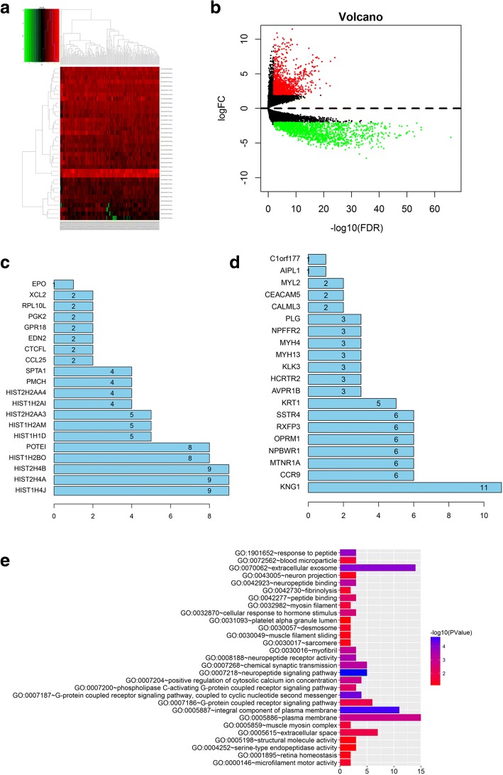
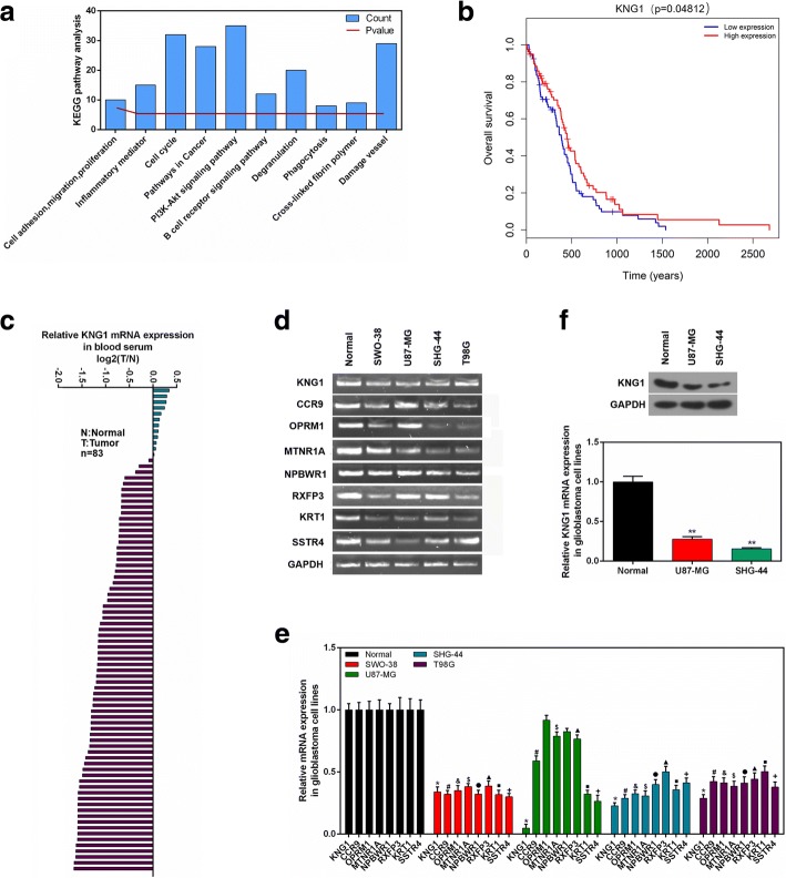
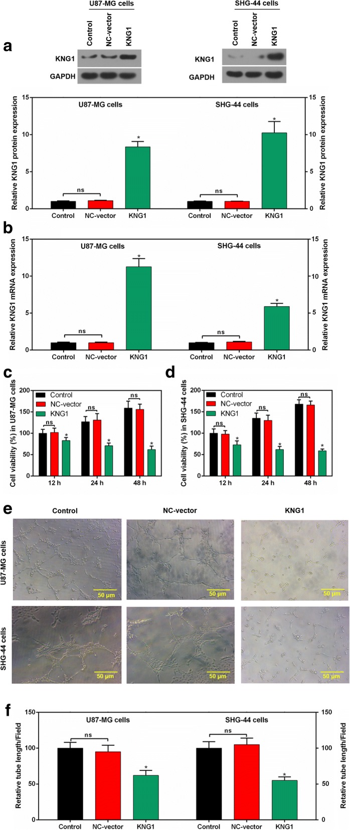
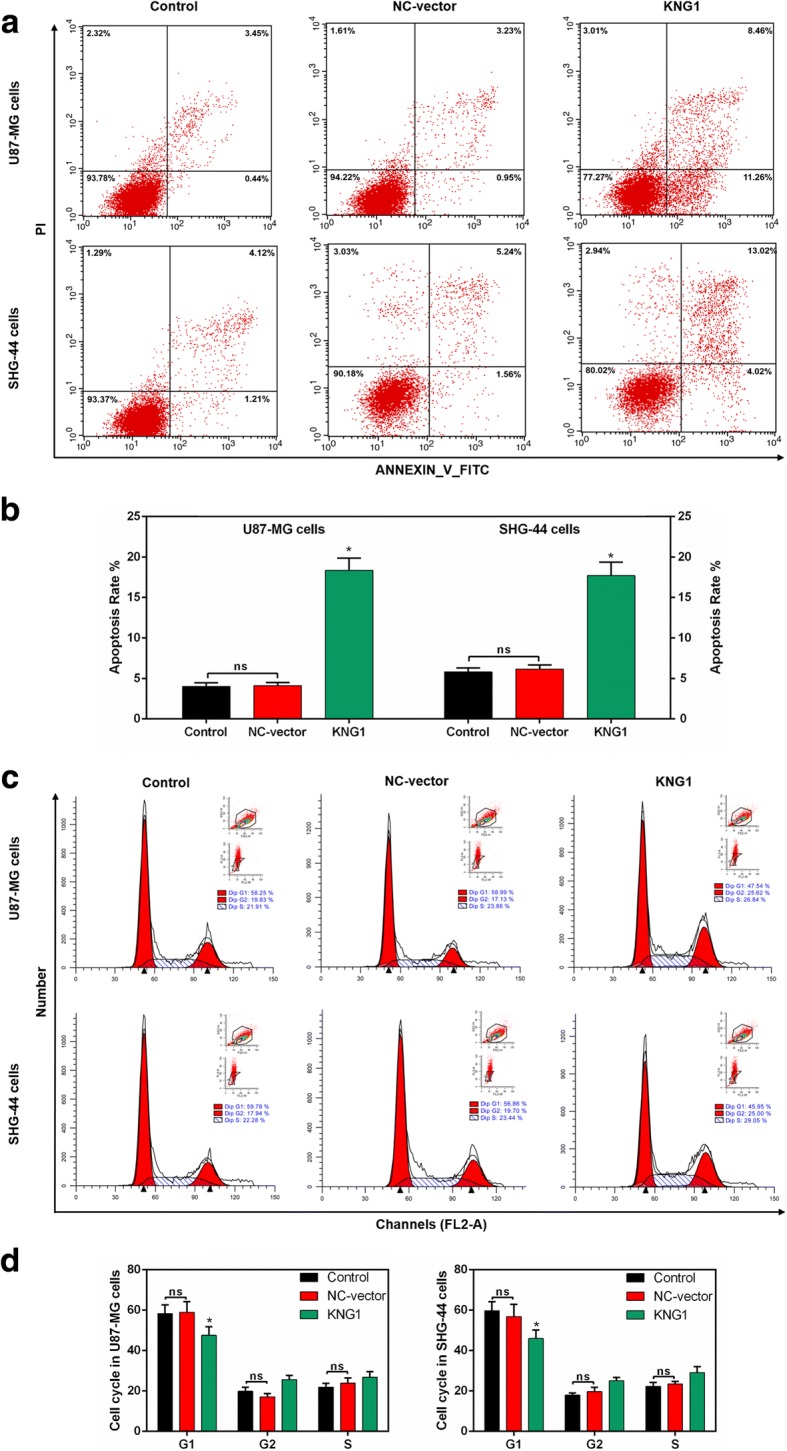
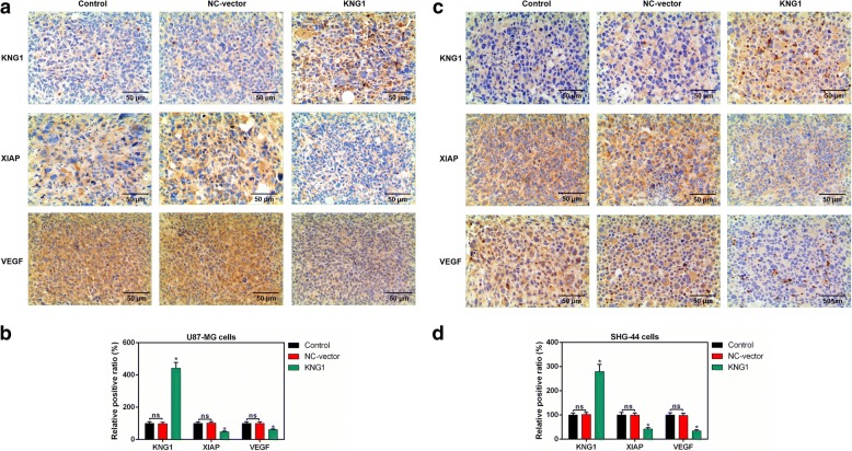
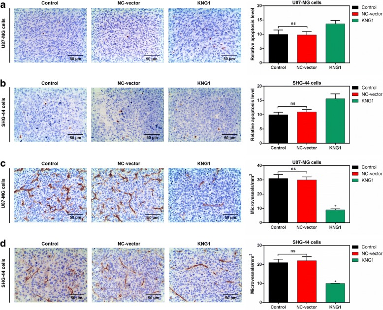
Methods: The differentially expressed genes (DEGs) were identified based on The Cancer Genome Atlas (TCGA) database. The KNG1-vector was transfected into the two glioma cells. The viability, apoptosis and cell cycle of glioma cells and microvessel density (MVD) were detected by cell counting kit-8 assay, flow cytometry and immunohistochemistry, respectively. The expression were measured by quantitative real-time PCR and Western blot, respectively. A tumor mouse model was established to determine apoptosis rate of brain tissue by terminal deoxynucleotidyl transfer-mediated dUTP nick end labeling (TUNEL) analysis.

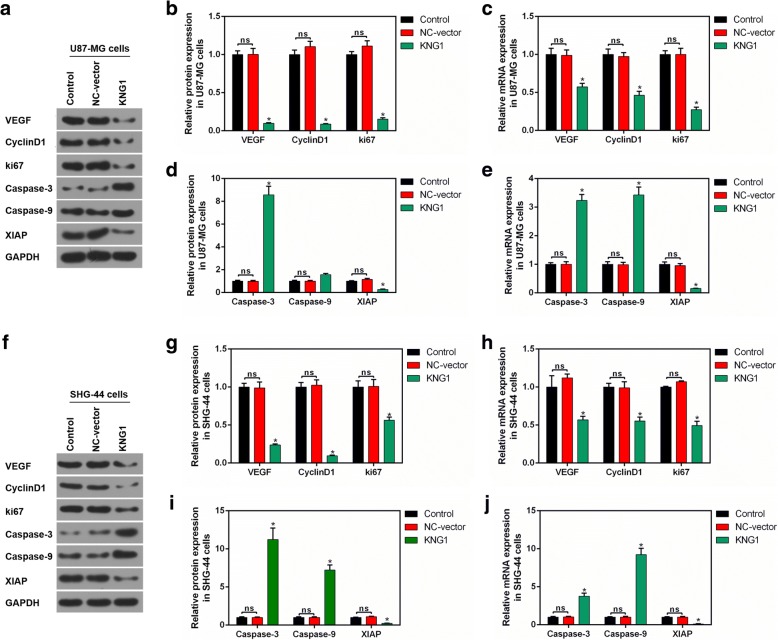
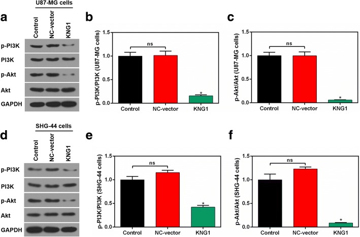
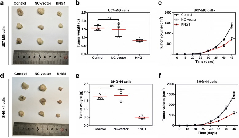
Results: KNG1 was identified as the core gene and lowly expressed in the glioma cells. Overexpression of KNG1 inhibited cell viability and angiogenesis of glioma cells. Overexpression of KNG1 promoted the apoptosis and G1 phase cell cycle arrest of glioma cells. Moreover, the expressions of VEGF, cyclinD1, ki67, caspase-3/9 and XIAP were regulated by overexpression of KNG1. In addition, overexpression of KNG1 inhibited the activity of PI3K/Akt. Furthermore, overexpression of KNG1 decreased the tumor growth and promoted the apoptosis of decreased by overexpression of KNG1 in vivo. .

Conclusions: Overexpression of KNG1 suppresses glioma progression by inhibiting the proliferation and promoting apoptosis of glioma cells, providing a therapeutic strategy for the malignant glioma.

Keywords: Angiogenesis; Apoptosis; Glioma; KNG1.


Conflict of interest statement

All procedures performed in studies involving human participants were in accordance with the ethical standards of the institutional and/or national research committee and with the 1964 Helsinki declaration and its later amendments or comparable ethical standards.

Not applicable.

The authors declare that they have no competing interests.

Springer Nature remains neutral with regard to jurisdictional claims in published maps and institutional affiliations.

Figures

Fig. 1
Fig. 1
Identification of core genes in screened differentially expressed genes (DEGs). a Heatmap of DEGs. DEMs with log2FC > 1 were red; DEMs with log2FC < − 1 were green. b Volcano plot of DEGs. DEGs with log2FC > 1.5 were expressed in red; DEGs with log2FC < − 1.5 were in green (P < 0.05). The down-regulated DEGs (c) and up-regulated DEGs (d) were identified. e The GO analysis of KNG1
Fig. 2
Fig. 2
Clinical analysis and cell verification. a The KEGG enrichment assay of KNG1. b Kaplan-Meier survival analysis for glioma patients with different expressions of KNG1. c The mRNA expression of KNG1 in serum from 69 human glioma patients and 14 normal patients. d The top 8 core genes detected by RT-PCR. e The expressions of top 8 core genes in four glioma cells. f The KNG1 was lowly expressed in the U87-MG and SHG-44 cells. P < 0.05, ★★P < 0.01
Fig. 3
Fig. 3
Overexpressed KNG1 inhibited the viability and angiogenesis of glioma cells. KNG1 was overexpressed in the glioma cells at protein (a) and mRNA levels (b). KNG1 overexpression inhibited the cell viability in a time-dependent manner in U87-MG (c) and SHG-44 cells (d). KNG1 overexpression suppressed the regenerative blood vessels (e) and reduced the length of blood vessel (f). The ns represents no significant difference; P < 0.05 versus control
Fig. 4
Fig. 4
Roles of KNG1 in the apoptosis and cell cycle. a Flow cytometry results of glioma cells. b The overexpressed KNG1 induced the apoptosis of U87-MG and SHG-44 cells. c-d Cell cycle analysis showed a decreased number of U87-MG and SHG-44 cells in G1 phase. The ns represents no significant difference; P < 0.05 versus control
Fig. 5
Fig. 5
Effect of KNG1 on the proteins associated with cell proliferation and apoptosis. a Protein levels of proliferation- and apoptosis-associated proteins were measured in U87-MG cells. b-c The expression of VEGF, CyclinD1 and ki67 were decreased by overexpressed KNG1. d-e The levels of caspase-3 and caspase-9 were enhanced while the XIAP expression was suppressed by overexpressed KNG1. f Changes of proliferation- and apoptosis-associated proteins were detected in SHG-44 cells. g-h The expression of VEGF, CyclinD1 and ki67 were inhibited by overexpressed KNG1. i-j The levels of caspase-3 and caspase-9 were increased while the XIAP expression was suppressed by overexpressed KNG1. The ns represents no significant difference; P < 0.05 versus control
Fig. 6
Fig. 6
Overexpressed KNG1 suppressed the activation of PI3K and Akt. a The levels of p-PI3K, PI3K, p-Akt and Akt by Western blot in U87-MG cells. The ratios of p-PI3K/PI3K (b) and p-Akt/Akt (c) were inhibited by KNG1 overexpression in U87-MG cells. d The levels of p-PI3K, PI3K, p-Akt and Akt by Western blot in SHG-44 cells. The ratios of p-PI3K/PI3K (e) and p-Akt/Akt (f) were inhibited by KNG1 overexpression in SHG-44 cells. The ns represents no significant difference; P < 0.05 versus control
Fig. 7
Fig. 7
KNG1 overexpression blocked tumorigenesis of glioma in vivo. a Representative images of xenograft tumors grown in nude mice injected with U87-MG cells. The weight (b) and volume (c) of tumors were inhibited by KNG1 overexpression. d Representative images of xenograft tumors grown in nude mice injected with SHG-44 cells. The weight (e) and volume (f) of tumors were also suppressed by KNG1 overexpression. The ns represents no significant difference; P < 0.05 versus control
Fig. 8
Fig. 8
Expressions of KNG1, VEGF and XIAP in brain tissues of mice. a The expression of KNG1, VEGF and XIAP in brain tissues of mice injected with U87-MG cells were measured by IHC. b The KNG1 level was increased while the expressions of VEGF and XIAP were decreased in brain tissues of mice injected with U87-MG cells. c The levels of KNG1, VEGF and XIAP in brain tissues of mice injected with SHG-44 cells. d The KNG1 level was increased while the expressions of VEGF and XIAP were decreased in brain tissues of mice injected with SHG-44 cells. The ns represents no significant difference; P < 0.05 versus control
Fig. 9
Fig. 9
Effects of KNG1 on the apoptosis and microvessles. KNG1 overexpression slightly increased the apoptosis of brain tissue in mice injected with U87-MG (a) and SHG-44 cells (b). However, KNG1 overexpression obviously inhibited the formation of microvessles in mice injected with U87-MG (c) and SHG-44 cells (d). The ns represents no significant difference; P < 0.05 versus control

References

    1. Onishi M, Date I. Angiogenesis and invasion in glioma. Brain Tumor Pathology. 2011;28(1):13–24. doi: 10.1007/s10014-010-0007-z. - DOI - PubMed
    1. Taylor LP. Diagnosis, treatment, and prognosis of glioma: five new things. Neurology. 2010;75(1):28–32. doi: 10.1212/WNL.0b013e3181fb3661. - DOI - PubMed
    1. Louis DN, Pomeroy SL, Cairncross JG. Focus on central nervous system neoplasia. Cancer Cell. 2002;1(2):125–128. doi: 10.1016/S1535-6108(02)00040-5. - DOI - PubMed
    1. Preusser M, Haberler C, Hainfellner JA. Malignant glioma: neuropathology and neurobiology. Wien Med Wochenschr. 2006;156(11–12):332–337. doi: 10.1007/s10354-006-0304-7. - DOI - PubMed
    1. Hardee ME, Zagzag D. Mechanisms of glioma-associated neovascularization. Am J Pathol. 2012;181(4):1126–1141. doi: 10.1016/j.ajpath.2012.06.030. - DOI - PMC - PubMed

MeSH terms