

Prenatal neural origins of infant motor development: Associations between fetal brain and infant motor development
- PMID: 30068433
- PMCID: PMC6261435
- DOI: 10.1017/S095457941800072X
Prenatal neural origins of infant motor development: Associations between fetal brain and infant motor development
Abstract
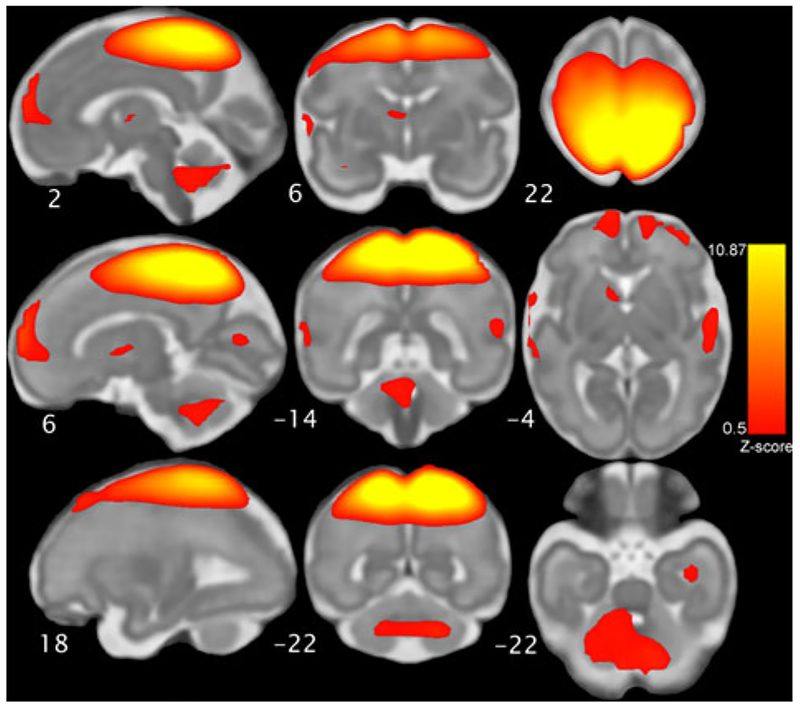
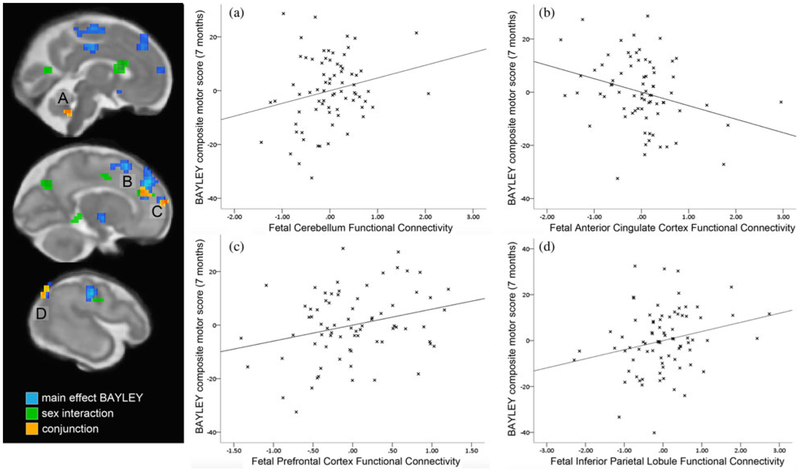
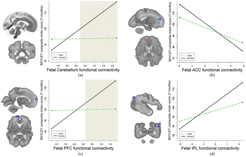
Functional circuits of the human brain emerge and change dramatically over the second half of gestation. It is possible that variation in neural functional system connectivity in utero predicts individual differences in infant behavioral development, but this possibility has yet to be examined. The current study examines the association between fetal sensorimotor brain system functional connectivity and infant postnatal motor ability. Resting-state functional connectivity data was obtained in 96 healthy human fetuses during the second and third trimesters of pregnancy. Infant motor ability was measured 7 months after birth using the Bayley Scales of Infant Development. Increased connectivity between the emerging motor network and regions of the prefrontal cortex, temporal lobes, posterior cingulate, and supplementary motor regions was observed in infants that showed more mature motor functions. In addition, females demonstrated stronger fetal-brain to infant-behavior associations. These observations extend prior longitudinal research back into prenatal brain development and raise exciting new ideas about the advent of risk and the ontogeny of early sex differences.
Figures
References
-
- Allen EA, Erhardt EB, Damaraju E, Gruner W, Segall JM, Silva RF, … Calhoun VD (2011). A baseline for the multivariate comparison of resting-state networks. Frontiers in System Neuroscience, 5, 2. doi:10.3389/fnsys.2011.00002 - DOI - PMC - PubMed
-
- Almli CR, Ball RH, & Wheeler ME (2001). Human fetal and neonatal movement patterns: Gender differences and fetal-to-neonatal continuity. Developmental Psychobiology, 38, 252–273. - PubMed
-
- Arca-Diaz G, Re TJ, Drottar M, Fortuno CR, De Macedo-Rodrigues K, Im K, … Grant PE (2017). Can cerebellar and brainstem apparent diffusion coefficient (ADC) values predict neuromotor outcome in term neonates with hypoxic-ischemic encephalopathy (HIE) treated with hypothermia? PLOS ONE, 12, e0178510. doi:10.1371/journal.pone.0178510 - DOI - PMC - PubMed
-
- Bale TL, & Epperson CN (2015). Sex differences and stress across the lifespan. Nature Neuroscience, 18. doi:10.1038/nn.4112 - DOI - PMC - PubMed
-
- Barnett MA, & Scaramella LV (2013). Mothers’ parenting and child sex differences in behavior problems among African American preschoolers. Journal of Family Psychology, 27, 773–783. doi:10.1037/a0033792 - DOI - PMC - PubMed
Publication types
MeSH terms
Grants and funding
LinkOut - more resources
Full Text Sources
Other Literature Sources
