. 2018 Sep 28;293(39):15084-15094.
doi: 10.1074/jbc.RA118.004564. Epub 2018 Aug 1.

The nature of the DNA substrate influences pre-catalytic conformational changes of DNA polymerase β

Affiliations

The nature of the DNA substrate influences pre-catalytic conformational changes of DNA polymerase β

Ji Huang et al. J Biol Chem. .

Abstract

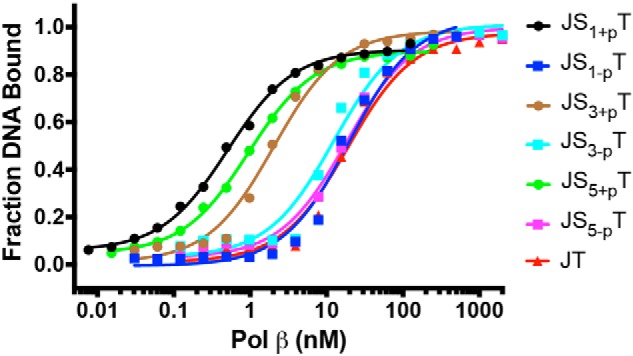
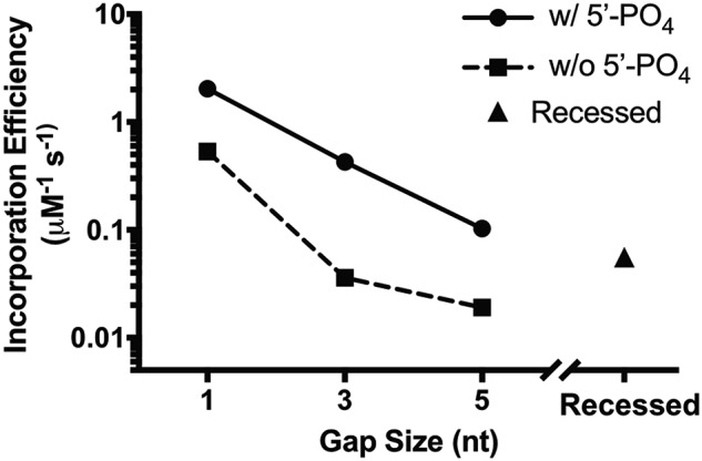
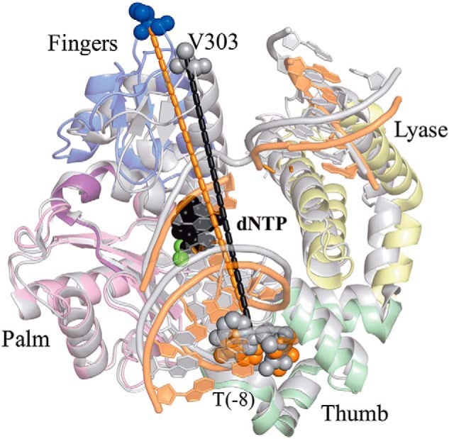
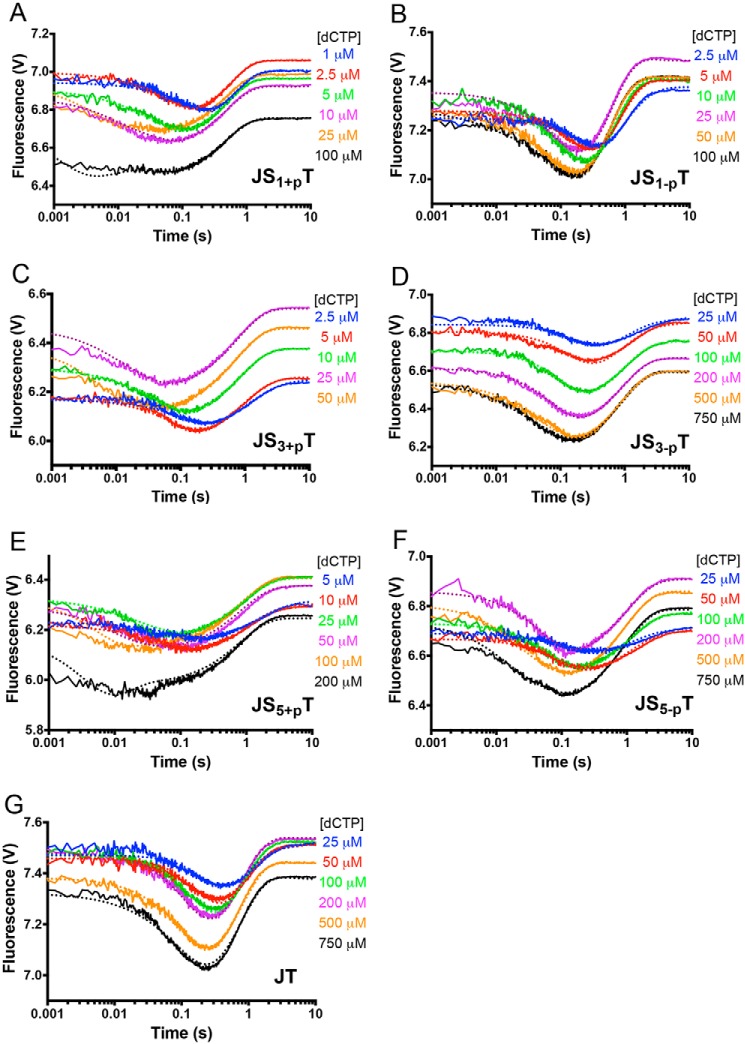
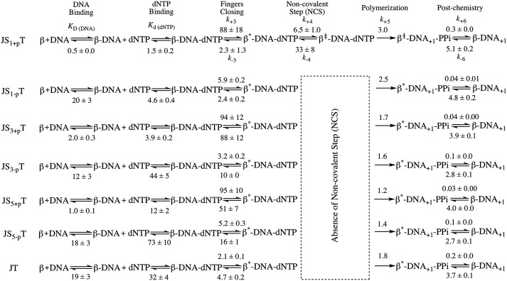
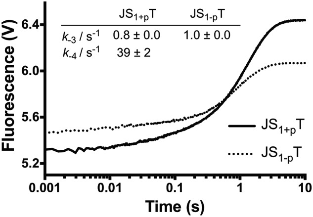
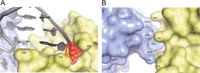
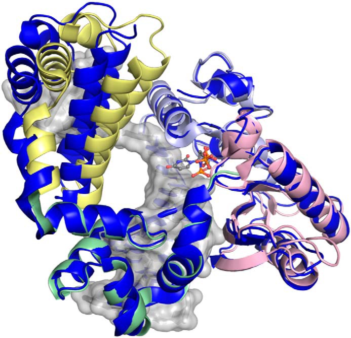
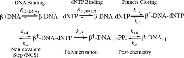
DNA polymerase β (Pol β) is essential for maintaining genomic integrity. During short-patch base excision repair (BER), Pol β incorporates a nucleotide into a single-gapped DNA substrate. Pol β may also function in long-patch BER, where the DNA substrate consists of larger gap sizes or 5'-modified downstream DNA. We have recently shown that Pol β fills small gaps in DNA during microhomology-mediated end-joining as part of a process that increases genomic diversity. Our previous results with single-nucleotide gapped DNA show that Pol β undergoes two pre-catalytic conformational changes upon binding to the correct nucleotide substrate. Here we use FRET to investigate nucleotide incorporation of Pol β with various DNA substrates. The results show that increasing the gap size influences the fingers closing step by increasing its reverse rate. However, the 5'-phosphate group has a more significant effect. The absence of the 5'-phosphate decreases the DNA binding affinity of Pol β and results in a conformationally more open binary complex. Moreover, upon addition of the correct nucleotide in the absence of 5'-phosphate, a slow fingers closing step is observed. Interestingly, either increasing the gap size or removing the 5'-phosphate group results in loss of the noncovalent step. Together, these results suggest that the character of the DNA substrate impacts the nature and rates of pre-catalytic conformational changes of Pol β. Our results also indicate that conformational changes are important for the fidelity of DNA synthesis by Pol β.

Keywords: 5′-phosphate; DNA polymerase; FRET; conformational change; crystal structure; gap size; kinetics; polymerase β.


Conflict of interest statement

The authors declare that they have no conflicts of interest with the contents of this article. The content is solely the responsibility of the authors and does not necessarily represent the official views of the National Institutes of Health

Figures

Figure 1.
Figure 1.
The 5′-phosphate group has a dominant effect on the binding of Pol β. DNA (0.1 nm) was incubated with varying amounts of Pol β (0.0076–2000 nm) in binding buffer for 15 min at 23 °C. Sigmoidal plots of the fraction of DNA bound versus the log of protein concentration show the differences for all seven DNA substrates. The binding affinities of Pol β to various DNA substrates are 0.5 ± 0.0 nm (JS1+pT), 20 ± 3 nm (JS1-pT), 2.0 ± 0.3 nm (JS3+pT), 12 ± 3 nm (JS3-pT), 1.0 ± 0.1 nm (JS5+pT), 18 ± 3 nm (JS5-pT), and 19 ± 3 nm (JT).
Figure 2.
Figure 2.
Effect of gap size and 5′-phosphate group on the correct nucleotide incorporation efficiency by Pol β.
Figure 3.
Figure 3.
The FRET system monitors the distance (r) change between the IAEDANS label on V303C of Pol β and the Dabcyl label on template DNA at the −8 position. Addition of the correct nucleotide induces a movement of the fingers domain, resulting in shortening of the distance (r) between the IAEDANS label on V303C (blue to gray) and the Dabcyl label on the template DNA at the −8 position. The PDB codes for ternary (gray tones) and binary (colored by domain) are 2FMS and 3ISB, respectively. Structures are superimposed via the palm domain.
Figure 4.
Figure 4.
FRET demonstrates that the binary complex of Pol β and DNA substrates with 5′-phosphate groups undergo faster fingers closing upon binding to the correct nucleotide. A–G, the fingers domain of Pol β closes in the presence of increasing dCTP concentrations for JS1+pT (A), JS1-pT (B), JS3+pT (C), JS3-pT (D), JS5+pT (E), JS5-pT (F), and JT (G) DNA. The nucleotide concentrations used are listed in each panel. The dotted lines from the KinTek Explorer modeling results overlay the experimentally determined FRET traces, shown as solid lines.
Scheme 1.
Scheme 1.
Kinetic scheme of Pol β with various DNA substrates. The noncovalent step is only present for JS1+pT DNA but not for other DNA substrates. The parameters are listed for each step. The overall fingers closing forward constant K3 = k+3/k−3 is calculated for each DNA substrate, and the values are 38, 1.1, 1.9, 2.4, 0.3, 0.3, and 0.4 for JS1+pT, JS3+pT, JS5+pT, JS1-pT, JS3-pT, JS5-pT, and JT, respectively. The unit for KD (DNA) is nanomolar and for Kd (dNTP) is micromolar. The units for fingers closing, the noncovalent step, and polymerization (both forward and reverse reaction) are per second. The unit for the post-chemistry step forward reaction is per second and for the reverse reaction is per micromolar per second.
Figure 5.
Figure 5.
Reverse fingers closing. We measured the reverse fingers closing rates by mixing preformed ternary complex and excess of binary complex, followed by monitoring the FRET signal as described under “Experimental procedures.” The data of JS1+pT were fit to a double exponential equation to generate a k−4 at 39 ± 2 s−1 and k−3 at 0.8 ± 0.0 s−1, where the rate of k−3 corresponds to 90% of the signal. The data of JS1-pT can only be fit with a single exponential equation to generate a k−3 at 1.0 ± 0.0 s−1.
Figure 6.
Figure 6.
A, the lyase domain of Pol β interacts with DNA in the ternary structure. Lys-35 and Lys-68 interact with the 5′-phosphate group on the downstream DNA. His-34 stacks with the nucleobase at the T+1 position of the template DNA. B, interactions between the lyase domain (yellow) and fingers domain (light blue) of Pol β in the ternary structure. The PDB code for structure is 2FMS (47).
Figure 7.
Figure 7.
Shown is a ternary complex of Pol β containing 1-nt gapped DNA with a 5′-phosphate group (PDB code 2FMS) (47), with each domain shown by color (lyase, yellow; thumb, green; palm, pink; fingers, light blue) superimposed with a recessed ternary structure, PDB code 2BPG) (6), shown in dark blue, with the only significant deviation in global conformation observed for the lyase domain.
None
None

References

    1. Gates K. S. (2009) An overview of chemical processes that damage cellular DNA: spontaneous hydrolysis, alkylation, and reactions with radicals. Chem. Res. Toxicol. 22, 1747–1760 10.1021/tx900242k - DOI - PMC - PubMed
    1. Cadet J., Douki T., and Ravanat J. L. (2010) Oxidatively generated base damage to cellular DNA. Free Radic. Biol. Med. 49, 9–21 10.1016/j.freeradbiomed.2010.03.025 - DOI - PubMed
    1. Barnes D. E., and Lindahl T. (2004) Repair and genetic consequences of endogenous DNA base damage in mammalian cells. Annu. Rev. Genet. 38, 445–476 10.1146/annurev.genet.38.072902.092448 - DOI - PubMed
    1. Matsumoto Y., and Kim K. (1995) Excision of deoxyribose phosphate residues by DNA polymerase β during DNA repair. Science 269, 699–702 10.1126/science.7624801 - DOI - PubMed
    1. Freudenthal B. D., Beard W. A., Shock D. D., and Wilson S. H. (2013) Observing a DNA polymerase choose right from wrong. Cell 154, 157–168 10.1016/j.cell.2013.05.048 - DOI - PMC - PubMed

Publication types

LinkOut - more resources