. 2018 Sep 28;293(39):14974-14988.
doi: 10.1074/jbc.RA118.003444. Epub 2018 Aug 1.

Small molecule-based targeting of TTD-A dimerization to control TFIIH transcriptional activity represents a potential strategy for anticancer therapy

Affiliations

Small molecule-based targeting of TTD-A dimerization to control TFIIH transcriptional activity represents a potential strategy for anticancer therapy

Virginie Gervais et al. J Biol Chem. .

Abstract

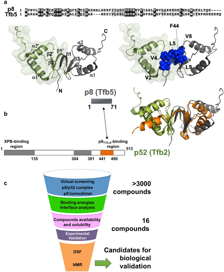
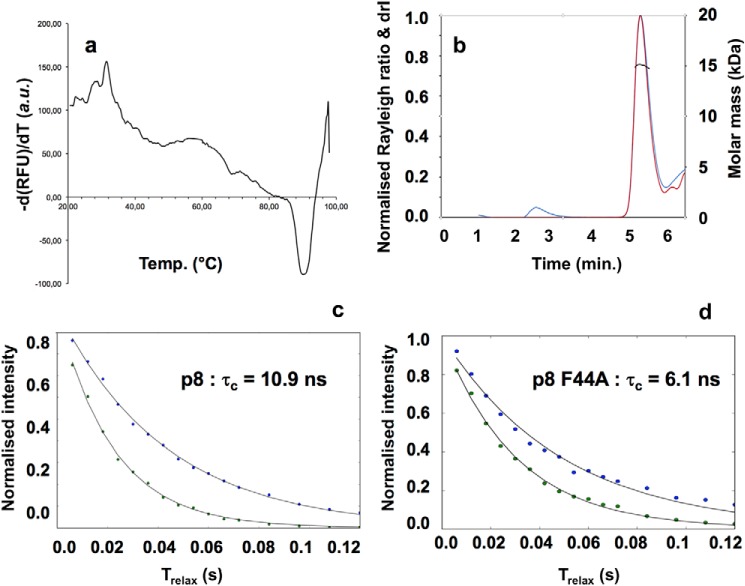
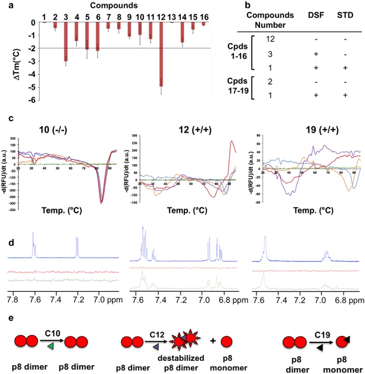
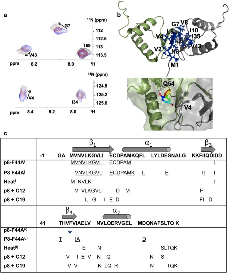
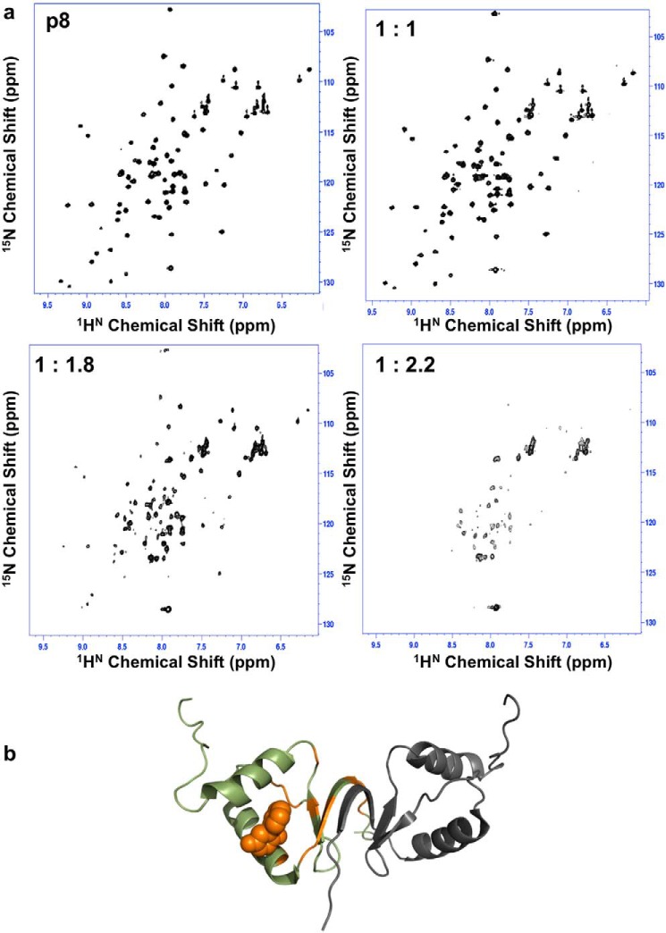
The human transcription factor TFIIH is a large complex composed of 10 subunits that form an intricate network of protein-protein interactions critical for regulating its transcriptional and DNA repair activities. The trichothiodystrophy group A protein (TTD-A or p8) is the smallest TFIIH subunit, shuttling between a free and a TFIIH-bound state. Its dimerization properties allow it to shift from a homodimeric state, in the absence of a functional partner, to a heterodimeric structure, enabling dynamic binding to TFIIH. Recruitment of p8 at TFIIH stabilizes the overall architecture of the complex, whereas p8's absence reduces its cellular steady-state concentration and consequently decreases basal transcription, highlighting that p8 dimerization may be an attractive target for down-regulating transcription in cancer cells. Here, using a combination of molecular dynamics simulations to study p8 conformational stability and a >3000-member library of chemical fragments, we identified small-molecule compounds that bind to the dimerization interface of p8 and provoke its destabilization, as assessed by biophysical studies. Using quantitative imaging of TFIIH in living mouse cells, we found that these molecules reduce the intracellular concentration of TFIIH and its transcriptional activity to levels similar to that observed in individuals with trichothiodystrophy owing to mutated TTD-A Our results provide a proof of concept of fragment-based drug discovery, demonstrating the utility of small molecules for targeting p8 dimerization to modulate the transcriptional machinery, an approach that may help inform further development in anticancer therapies.

Keywords: DNA transcription; GTF2H5; STD-NMR; TFIIH; cancer; drug screening; nuclear magnetic resonance (NMR); protein-protein interaction; quantitative imaging; transcriptional regulation.


Conflict of interest statement

The authors declare that they have no conflicts of interest with the contents of this article

Figures

Figure 1.
Figure 1.
Structure of the TFIIH p8 subunit and protein–protein interface. a (top), sequence alignment of human p8 and yeast Tfb5. Left, solution structure of the human p8 homodimer (PDB code 2JNJ). The two monomers are depicted as green and gray ribbon, and their secondary structure elements are indicated. Right, the same view highlighting as blue spheres the residues located at the interface between the two monomers. b, schematic organization of p8 (residues 1–71, Tfb5 in yeast) and p52 (residues 1–513, Tfb2 in yeast) proteins. Gray boxes in p52 indicate the two regions that participate in the binding with the XPB helicase, whereas the orange box indicates the region of interaction with p8. Right, structure of p8 with the same color code as in a was superimposed onto the crystal structure of the Tfb5–Tfb2C complex (Tfb5 in green and Tfb2 in orange). Both structures could be superimposed for 67 atoms with a root mean square deviation value of 5.8 Å. c, flow chart showing the different steps of the screening process used in our work.
Figure 2.
Figure 2.
p8 is a dimer in solution. a, fluorescence signal (a.u., arbitrary units) as a function of temperature giving unfolding temperature of p8 in 50 mm Tris-HCl, 150 mm NaCl, 0.25 mm TCEP at pH 7.5, indicating high thermal stability (Tm ∼88 °C). The presence of 1 and 5% DMSO was found to leave this plot unaffected. b, elution profile obtained by SEC-MALS. The traces of the light scattering (blue), differential refractive index (red), and MALS-derived molar masses (black) are shown. The experimental SEC-MALS experiments gave a mean molar mass of 15,530 ± 120 g/mol−1, confirming the presence of homodimer species in solution (the calculated molecular mass of p8 is 8053.4 Da). c, NMR determination of the global rotational correlation times, at T = 298 K, for p8 and p8Phe44A (TRACT experiments (17)). The normalized intensity decay of the two 15N doublet coherences is plotted as a function of the relaxation delay. Fitting the data to mono-exponential functions returned the relaxation rate constants for α and β spin states. The difference between the two relaxation rates gave a global rotational correlation time τc of 10.9 ns (R = 23.3 Hz; R = 45.6 Hz) for p8, consistent with a dimer in solution. The τc value was significantly reduced in the case of p8Phe44A (6.1 ns; R = 20.3 Hz; R = 32.8 Hz), which is consistent with the expected value (5.7 ns) for a monomeric protein at this temperature (27).
Figure 3.
Figure 3.
Evaluation of potential binders using thermal shift assays and NMR. a, histogram illustrating the ligand-induced changes in p8 thermal stability. The ΔTm values indicate the resulting shifts compared with the Tm signal of the free protein (around 90 °C; see Fig. 2a). DSF experiments were performed with a final protein concentration of 70 μm and final ligand concentration of 0.5 mm, respectively. The error bars are based at least on triplicate experiments. b, table summarizing the results obtained by DSF and STD for the 19 compounds tested (compound structures are given in Table 1). c, DSF melting curves of p8 dimer upon the addition of compounds 10 (left), 12 (middle), and 19 (right). Increasing concentrations of compounds (0.1 (orange), 0.25 (purple), and 0.5 mm (red)) were added to protein samples (70–90 μm) in buffer consisting of 50 mm Tris-HCl, 150 mm NaCl, and 0.25 mm TCEP at pH 7.5. Green and blue lines indicate DSF curves obtained for the compounds (0.5 mm) and p8 alone, respectively. d, 1H NMR spectra of aromatic regions of the compounds (blue) and STD spectra recorded for the compounds in the absence of protein (red) and in the presence of 10 μm of protein (green). The compounds were present at 1 mm concentration in phosphate buffer. The broader lines observed for compound 19 may arise from the formation of a covalent adduct with p8 (see Figs. S3 and S4). e, schematic drawing illustrating the effect of the three compounds 10, 12, and 19 on p8 dissociation; compound 10 has no effect, compound 12 leads to partial dissociation of the p8 homodimer, and compound 19 promotes complete dissociation leading to monomeric species in solution. At high protein/ligand ratios (>10) and upon long incubation times, compound 19 can form a covalent adduct with p8 that also destabilizes p8 (see Figs. S3 and S4).
Figure 4.
Figure 4.
Compound 12 binds to the homodimer interface. a, selected view of the 1H-15N HSQC spectrum recorded for the homodimer of p8, illustrating chemical shift changes for residues Val4, Gly7, and Val43 at the interface of the dimer upon the addition of compound 12. b, cartoon representation of the homodimer structure of p8, indicating the residues exhibiting chemical shift perturbation (in blue). The two monomers are shown as gray and green ribbon representations. A representative docking pose of compound 12 indicates hydrogen bonding with Val4 from one subunit and Gln54 from the other subunit of the p8 homodimer. c, table summarizing the residues of p8 whose resonances are perturbed either in the Phe44 mutant (underlined residues; p8-F44A(i) (7) and p8-F44A(j) (our work)) or by heat(i) (7) or upon the addition of compounds 12 and 19 to the WT protein. Secondary structures are indicated above the p8 sequence.
Figure 5.
Figure 5.
Partial unfolding of p8 in the presence of compound 19. a, 1H-15N HSQC spectra recorded for p8 in the presence of increasing concentrations of compound 19, revealing gradual alteration of the quality of NMR spectra until partial protein denaturation for protein/ligand ratios exceeding 1:1.8. b, cartoon representation of the homodimer structure of p8 mapping positions of residues exhibiting severe line-broadening until the limit of NMR detection (orange) in the presence of compound 19, shown as an orange sphere (predicted binding pose obtained from docking).
Figure 6.
Figure 6.
Stability of TFIIH in living cells. a and b, Western blotting of the XPB subunit of TFIIH in ES cells (a) and chondrocytes (b) from the XPB-YFP mouse model (ES-XY and C-XY, respectively) after 24, 48, and 72 h of treatment with the protein synthesis inhibitor cycloheximide (133 μg/ml; NT, untreated; D, DMSO; E, ethanol). c, fluorescent nuclei of ES-XY cells at the 0-, 5-, 15-, and 20-h time points after treatment with different concentrations of cycloheximide (Scale bar: 10 μm.).
Figure 7.
Figure 7.
Reduction of TFIIH steady-state and transcriptional activity. a, typical examples of XPB-YFP–expressing cells, 5 and 10 days after continued treatment with compounds 10 and 12 at 1 μm concentration. b, results for the quantification of TFIIH levels via measurement of total XPB-YFP intensities in the nucleus of cells treated with 3 μm compounds 10, 12, and 19 after 10 days of treatment. c, results for the quantification of transcriptional activity in cells treated 10 days with compounds 10, 12, and 19 and in XPB-YFPTTDA−/− cells. Error bars, S.E. ***, p < 0.001, Student's t test (two tailed, assuming unequal variances).

References

    1. Luo J., Cimermancic P., Viswanath S., Ebmeier C. C., Kim B., Dehecq M., Raman V., Greenberg C. H., Pellarin R., Sali A., Taatjes D. J., Hahn S., and Ranish J. (2015) Architecture of the human and yeast general transcription and DNA repair factor TFIIH. Mol. Cell 59, 794–806 10.1016/j.molcel.2015.07.016 - DOI - PMC - PubMed
    1. Greber B. J., Nguyen T. H. D., Fang J., Afonine P. V., Adams P. D., and Nogales E. (2017) The cryo-electron microscopy structure of human transcription factor IIH. Nature 549, 414–417 10.1038/nature23903 - DOI - PMC - PubMed
    1. Theil A. F., Nonnekens J., Wijgers N., Vermeulen W., and Giglia-Mari G. (2011) Slowly progressing nucleotide excision repair in trichothiodystrophy group A patient fibroblasts. Mol. Cell Biol. 31, 3630–3638 10.1128/MCB.01462-10 - DOI - PMC - PubMed
    1. Giglia-Mari G., Coin F., Ranish J. A., Hoogstraten D., Theil A., Wijgers N., Jaspers N. G. J., Raams A., Argentini M., van der Spek P. J., Botta E., Stefanini M., Egly J. M., Aebersold R., Hoeijmakers J. H. J., and Vermeulen W. (2004) A new, tenth subunit of TFIIH is responsible for the DNA repair syndrome trichothiodystrophy group A. Nat. Genet. 36, 714–719 10.1038/ng1387 - DOI - PubMed
    1. Coin F., Proietti De Santis L., Nardo T., Zlobinskaya O., Stefanini M., and Egly J. M. (2006) P8/TTD-A as a repair-specific TFIIH subunit. Mol. Cell 21, 215–226 10.1016/j.molcel.2005.10.024 - DOI - PubMed

Publication types

MeSH terms

LinkOut - more resources