. 2018 Sep 21;293(38):14678-14688.
doi: 10.1074/jbc.RA118.004343. Epub 2018 Aug 1.

Agonistic β-Klotho antibody mimics fibroblast growth factor 21 (FGF21) functions

Affiliations

Agonistic β-Klotho antibody mimics fibroblast growth factor 21 (FGF21) functions

Xiaoshan Min et al. J Biol Chem. .

Abstract

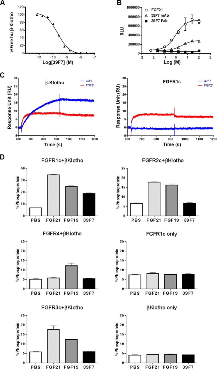
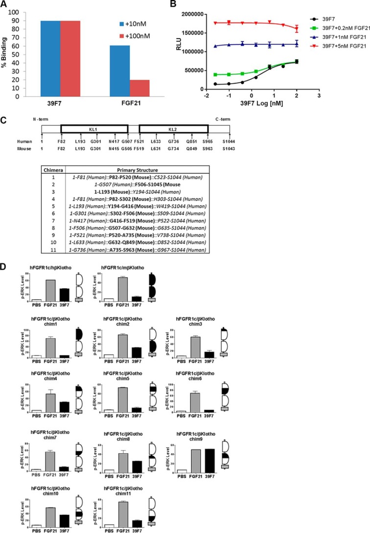
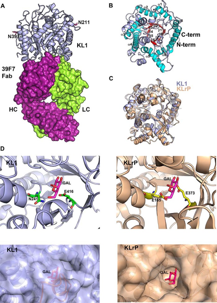
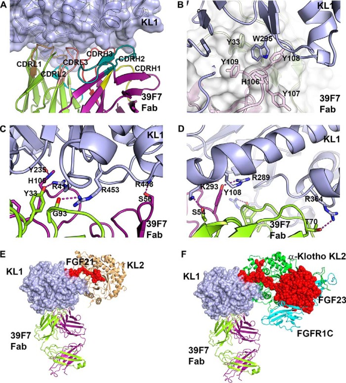
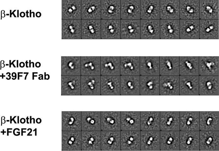
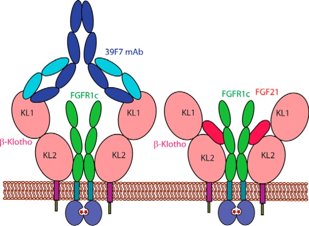
Fibroblast growth factor 21 (FGF21), an endocrine hormone in the FGF family, plays a critical role in regulating metabolic homeostasis and has emerged as a therapeutic target for metabolic diseases, including Type 2 diabetes mellitus. FGF21 functions through a receptor complex that consists of an FGF receptor (FGFR) and a co-receptor β-Klotho. Here, we identify and biochemically and structurally characterize 39F7, a high-affinity agonistic monoclonal antibody (mAb) against β-Klotho that mimics FGF21 function. The co-crystal structure of β-Klotho KL1 domain in complex with 39F7 Fab revealed that the recognition of 39F7 is centered on Trp-295 of β-Klotho in a FGF21 noncompetitive manner. KL1 adopts a (β/α)8 TIM barrel fold which resembles that of β-glycosylceramidase, but lacks molecular features for enzymatic activity, suggesting that KL1 functions as a scaffold protein instead. In vitro characterization demonstrated that, although 39F7 does not compete with FGF21, it is specific for β-Klotho/FGFR1c activation. Furthermore, the agonistic activity of 39F7 required the full IgG molecule to be bivalent, suggesting that 39F7 functions by promoting receptor/co-receptor dimerization. Supported by negative stain EM analysis of full-length β-Klotho, we propose a molecular model wherein the agonistic antibody 39F7 acts in a β-Klotho- and FGFR1c-dependent manner, mimicking FGF21 activity. More importantly, 39F7 offers promising therapeutic potential in the axis of FGF21 signaling as an antibody therapy alternative to FGF21 analogs for treatment of metabolic diseases.

Keywords: agonist; antibody; binding; crystallography; electron microscopy (EM); fibroblast growth factor (FGF); metabolism; β-Klotho.


Conflict of interest statement

All authors are employees of Amgen

Figures

Figure 1.
Figure 1.
Biochemical characterization of 39F7. A, binding affinity of 39F7 to human β-Klotho by solution equilibrium assay on Biacore. Human β-Klotho at 10 nm was premixed with 39F7, and the binding of free β-Klotho was detected by injecting over immobilized 39F7 surface on Biacore T200. Shown here, binding of 10 nm β-Klotho alone as 100%, the relative binding of β-Klotho in the mix with 39F7 was plotted versus antibody concentration. B, FGF21, 39F7, or 39F7 Fab induces luciferase activity in an FGF21-responsive reporter cell line. Luciferase activity is measured in relative luminescence units (RLU). C, binding sensorgram of 39F7 to human β-Klotho and FGFR1c on Biacore. Human β-Klotho and FGFR1c were captured at an approximate density of 40 RU and 60 RU on an anti-His antibody surface. 39F7 (blue) and FGF21 (red) at 100 nm and 500 nm were injected over captured β-Klotho and FGFR1c. D, 39F7 requires FGFR1c and β-Klotho to induce signaling as measured by ERK phosphorylation in L6 cells. L6 cells were selected for this characterization, as they do not express any endogenous FGF receptors. Data are expressed as percent ERK protein that is phosphorylated, as measured by Meso Scale Discovery (MSD) assay.
Figure 2.
Figure 2.
39F7 binds to β-Klotho KL1 domain and does not compete with FGF21 binding to the co-receptor. A, competition of 39F7 with FGF21 on the binding to β-Klotho. Human β-Klotho at 10 nm was premixed with 39F7 or FGF21, and the binding of free β-Klotho was detected by injecting over immobilized FGF21 on Biacore T200. Using binding of 10 nm β-Klotho alone as 100%, the relative binding of β-Klotho premixed with 39F7 or FGF21 at different concentrations, 10 nm (blue) or 100 nm (red), was plotted. The data shown are representative of multiple repeats. B, the 39F7-induced luciferase reporter activity was tested in the absence or presence of different concentrations of FGF21. C and D, a series of human–mouse β-Klotho chimeras was designed (C) and co-transfected into L6 cells with human FGFR1c, and signaling was measured by ERK phosphorylation (D). The β-Klotho chimeras are represented by schematics next to the pERK histograms, with murine sequence indicated as black and human sequence represented as white. The plasma membrane is indicated in gray.
Figure 3.
Figure 3.
Structure of 39F7-KL1 complex. A, overall structure of 39F7 Fab/KL1 complex. KL1 domain is shown in cartoon representation and colored in light blue. 39F7 Fab is shown in surface representation and colored in purple and green for heavy chain and light chain, respectively. B, structure of KL1. KL1 is shown in cartoon representation and is colored by secondary structure: red for β-strand and cyan for α-helix in the TIM barrel fold and light blue for loop and additional helices not belonging to the TIM barrel fold. C, overlay of KL1 domain with KLrP protein (PDB ID: 2E9L). KL1 domain is colored in blue and KLrP protein is colored in wheat. D, comparison of the active site of KLrP and KL1 domain. Top panels: the KLrP protein is shown in wheat cartoon and the active site residues are shown as yellow sticks. Galactose is shown as magenta sticks. KL1 domain protein is shown as light blue cartoon and the corresponding active site residues are shown as green sticks. A modeled glucose molecule is shown in the KL1 active site in magenta sticks. Bottom panels: KLrP protein is shown as wheat surface and the KL1 domain protein is shown as light blue surface. Galactose molecule in KLrP and the modeled galactose molecule in KL1 are shown as magenta sticks. The peptide from 374 to 381 in KL1 is colored in orange.
Figure 4.
Figure 4.
Interactions between 39F7 Fab and KL1 domain. A, the interface between KL1 and 39F7 Fab. KL1 protein is colored in light blue. 39F7 Fab is colored in purple for heavy chain and green for light chain. The six CDR loops are colored in the following order: CDRH1, yellow; CDRH2, green; CDRH3, red; CDRL1, dark yellow; CDRL2, cyan; CDRL3, pink. B, detailed hydrophobic interactions at the interface. C and D, H-bond interactions at the interface. KL1 domain is shown as light blue cartoon. 39F7 Fab is shown as white surface representation. Selected KL1 residues and 39F7 Fab residues are shown in sticks. E, structure overlay of β-Klotho KL1 domain in complex with 39F7 Fab to full-length β-Klotho in complex with FGF21 C-terminal peptide (PDB ID:5VAQ). KL1 domain is shown as blue surface. The 39F7 Fab is shown as green cartoon for light chain and purple cartoon for heavy chain. FGF21 is shown as red sphere and KL2 domain is shown as wheat cartoon. F, structure overlay of β-Klotho KL1 domain in complex with 39F7 Fab to full-length α-Klotho in complex with FGF23 and FGFR1 (PDB ID:5W21). The KL1 domain is shown as blue surface. The 39F7 Fab is shown as green cartoon for light chain and purple cartoon for heavy chain. FGF23 is shown as red sphere. The KL2 domain is shown as green cartoon. FGFR1C is shown as cyan cartoon.
Figure 5.
Figure 5.
EM characterization of β-Klotho and its interactions with 39F7 antibody and ligand FGF21 in solution. 2D class averages are shown for full-length β-Klotho (top panel, labeled KL), β-Klotho + 39F7 Fab (middle panel, labeled KL + Fab), and β-Klotho + FGF21 (bottom panel, labeled KL + FGF21).
Figure 6.
Figure 6.
Model of the mechanism of action for 39F7 antibody activation of β-Klotho and FGFR1c.

References

    1. Itoh N., and Ornitz D. M. (2004) Evolution of the Fgf and Fgfr gene families. Trends Genet. 20, 563–569 10.1016/j.tig.2004.08.007 - DOI - PubMed
    1. Beenken A., and Mohammadi M. (2009) The FGF family: Biology, pathophysiology and therapy. Nat. Rev. Drug Discov. 8, 235–253 10.1038/nrd2792 - DOI - PMC - PubMed
    1. Zhang J., and Li Y. (2016) Therapeutic uses of FGFs. Semin. Cell Dev. Biol. 53, 144–154 10.1016/j.semcdb.2015.09.007 - DOI - PubMed
    1. Degirolamo C., Sabbà C., and Moschetta A. (2016) Therapeutic potential of the endocrine fibroblast growth factors FGF19, FGF21 and FGF23. Nat. Rev. Drug Discov. 15, 51–69 10.1038/nrd.2015.9 - DOI - PubMed
    1. Kuro-o M., Matsumura Y., Aizawa H., Kawaguchi H., Suga T., Utsugi T., Ohyama Y., Kurabayashi M., Kaname T., Kume E., Iwasaki H., Iida A., Shiraki-Iida T., Nishikawa S., Nagai R., and Nabeshima Y. I. (1997) Mutation of the mouse klotho gene leads to a syndrome resembling ageing. Nature 390, 45–51 10.1038/36285 - DOI - PubMed

Publication types