. 2018 Dec;37(50):6414-6424.
doi: 10.1038/s41388-018-0419-5. Epub 2018 Aug 1.

Hippo signaling dysfunction induces cancer cell addiction to YAP

Affiliations

Hippo signaling dysfunction induces cancer cell addiction to YAP

Han Han et al. Oncogene. 2018 Dec.

Abstract

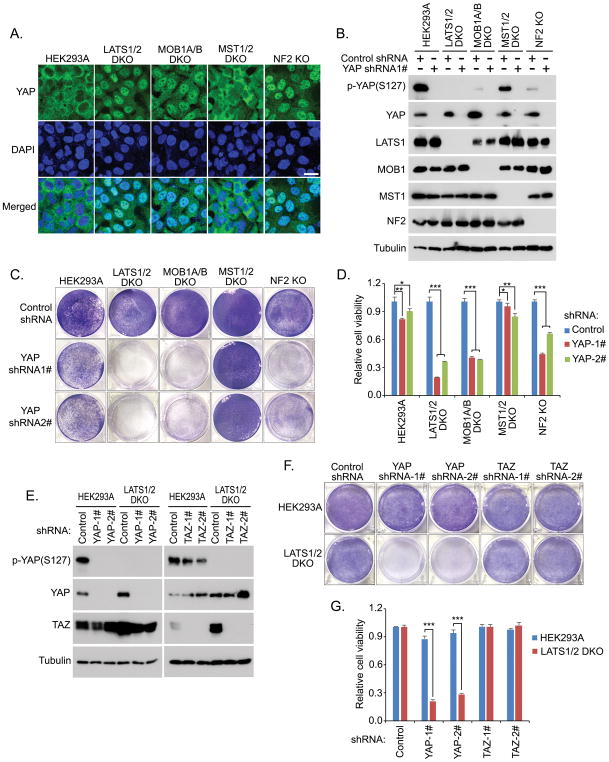
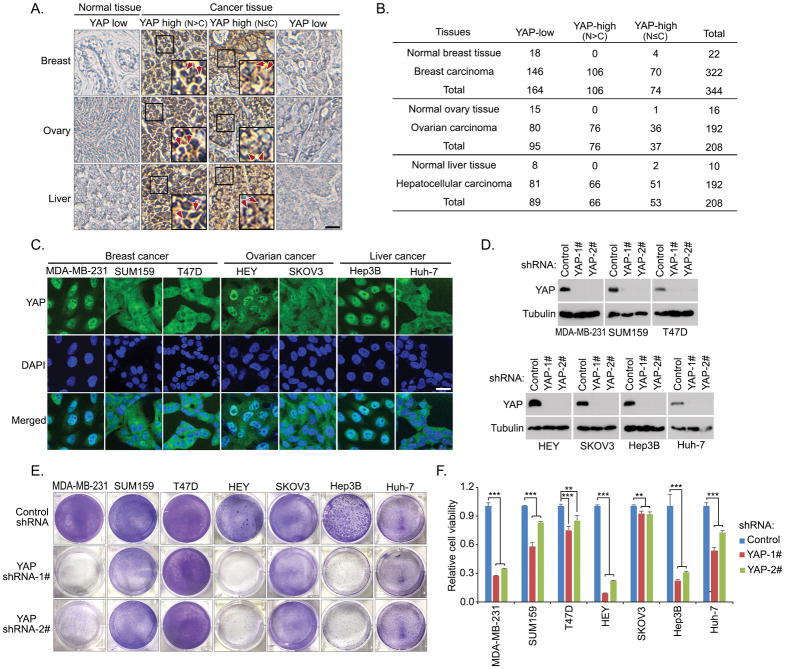
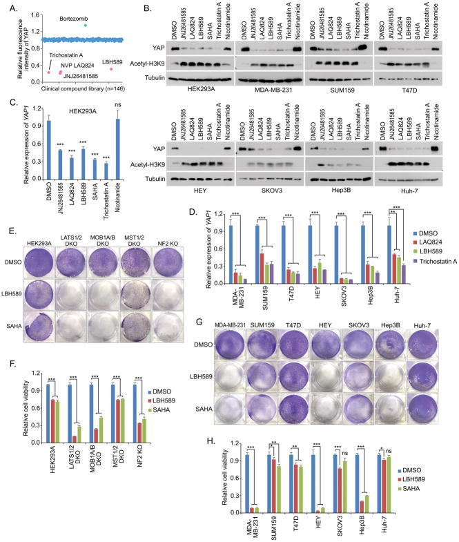
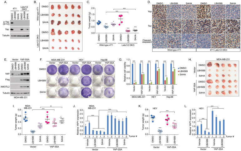
Over the past decades, the Hippo has been established as a crucial pathway involved in organ size control and cancer suppression. Dysregulation of Hippo signaling and hyperactivation of its downstream effector YAP are frequently associated with various human cancers. However, the underlying significance of such YAP activation in cancer development and therapy has not been fully characterized. In this study, we reported that the Hippo signaling deficiency can lead to a YAP-dependent oncogene addiction for cancer cells. Through a clinical compound library screen, we identified histone deacetylase (HDAC) inhibitors as putative inhibitors to suppress YAP expression. Importantly, HDAC inhibitors specifically targeted the viability and xenograft tumor growth for the cancer cells in which YAP is constitutively active. Taken together, our results not only establish an active YAP-induced oncogene addiction in cancer cells, but also lay the foundation to develop targeted therapies for the cancers with Hippo dysfunction and YAP activation.


Conflict of interest statement

Conflict of interest

The authors declare no competing conflict of interests.

Figures

Figure 1
Figure 1. Hippo signaling deficiency induces the cell addiction to YAP. (See also Figure S1)
(A) YAP is activated and accumulated in the nucleus of the LATS1/2 DKO, MOB1A/B DKO and NF2 KO cells. Loss of MST1/2 does not affect YAP cellular localization. Nucleus was visualized by DAPI. Scale bar, 20 μm. (B) Downregulation of YAP in the Hippo component KO cells. Western blotting was performed with indicated antibodies (C and D) Loss of YAP specifically suppressed the viability of the LATS1/2 DKO, MOB1A/B DKO and NF2 KO cells. Cell viability was visualized by crystal violet staining (C) and quantified (mean ± s.d., n = 3 biological replicates) (D). * p < 0.05, ** p < 0.01, *** p < 0.001. (E–G) Loss of YAP but not TAZ suppressed the LATS1/2 DKO cell viability. shRNA-mediated downregulation of YAP and TAZ was confirmed by Western blot in both wild-type HEK293A and LATS1/2 DKO cells (E). Cell viability was visualized by crystal violet staining (F) and quantified (mean ± s.d., n = 3 biological replicates) (G). *** p < 0.001.
Figure 2
Figure 2. Cancer cells with the active YAP exhibit the YAP dependence
(A and B) Immunohistochemical staining of YAP were performed in breast cancer, ovarian cancer and liver cancer tissue microarrays. Brown staining indicates positive immunoreactivity (A). Scale bar, 40 μm. The box region is twice enlarged. Arrows indicated nuclear staining of YAP. Correlation analysis of YAP expression/localization in the indicated human normal and tumor samples are shown as tables (B). (C) YAP is activated and accumulated in the nuclei of a group of cancer cell lines. YAP localization in each cancer cell was examined by immunofluorescence. Nucleus was visualized by DAPI. Scale bar, 20 μm. (DF) Loss of YAP specifically suppressed the viability of the cancer cells with YAP dominantly localized in the nucleus. shRNA-mediated downregulation of YAP was confirmed by Western blot in the indicated cancer cells (D). Cell viability was visualized by crystal violet staining (E) and quantified (mean ± s.d., n = 3 biological replicates) (F). ** p < 0.01, *** p < 0.001.
Figure 3
Figure 3. HDAC inhibitors suppress the YAP gene expression and the YAP-dependent cell viability. (See also Figure S2)
(A) A group of HDAC inhibitors were identified to suppress YAP expression. A clinical compound library (with 146 compounds) screen was performed in MDA-MB-231 cells. YAP immunofluorescent staining was performed and its relative fluorescent intensity was calculated. Several identified hits that affect YAP fluorescent intensity were indicated. (B) HDAC inhibitors suppressed the YAP protein expression. Western blotting was performed with the indicated antibodies. Cells were treated for 24 hours with the indicated compounds (0.5 μM JNJ26481585, 0.5 μM LAQ824, 0.5 μM LBH589, 10 μM SAHA, 10 μM Trichostatin A, and 100 μM Nicotinamide). (C and D) YAP gene transcription is suppressed by HDAC inhibitors. YAP gene transcription (mean ± s.d., n = 3 biological replicates) was examined in both HEK293A (C) and the indicated cancer cells (D) after 24 hour-treatment by the indicated compounds. ** p < 0.01, *** p < 0.001, ns, no significance. (E and F) HDAC inhibitors dramatically targeted the viability of the LATS1/2 DKO, MOB1A/B and NF2 KO cells compared to that of the wild-type and MST1/2 DKO cells. Cell viability was visualized by crystal violet staining (E) and quantified (mean ± s.d., n = 3 biological replicates) (F). *** p < 0.001. (G and H) HDAC inhibitors dramatically targeted the viability of the cancer cells with the active YAP addiction. Cell viability was visualized by crystal violet staining (G) and quantified (mean ± s.d., n = 3 biological replicates) (H). * p < 0.05, ** p < 0.01, *** p < 0.001, ns, no significance.
Figure 4
Figure 4. Hippo deficiency induces the tumor vulnerability to HDAC inhibitors
(A) HDAC inhibitors suppressed Yap expression in both wild-type 4T1 and Lats1/2 DKO 4T1 cells. Western blotting was performed with the indicated antibodies. (B and C) Loss of Lats1/2 sensitized the 4T1-derived xenograft tumors to HDAC inhibitors. Wild-type 4T1or Lats1/2 DKO 4T1cells were subjected to the xenograft tumor assay and treated with LBH589 and SAHA. Xenograft tumors are shown in (B) and the tumor weight was quantified (n = 5 mice, mean ± s.d.) (C). * p < 0.05, *** p < 0.001. (D) Immunohistochemistry was performed with the indicated antibodies in the HDAC inhibitor-treated wild-type 4T1 and Lats1/2 DKO 4T1 xenograft tumors. Scale bar, 40 μm. (E) HDAC inhibitors failed to suppress the expression of exogenously expressed YAP-5SA. Western blotting was performed with the indicated antibodies. (F and G) Overexpression of the constitutively active YAP-5SA mutant rescued the viability of cells with the active YAP addiction under the treatment of HDAC inhibitors. Control or YAP-5SA mutant-transfected MDA-MB-231, HEY and Hep3B cells were treated with LBH589 and SAHA. Cell viability was visualized by crystal violet staining (F) and quantified (mean ± s.d., n = 3 biological replicates) (G). (H–J) Overexpression of the constitutively active YAP-5SA mutant rescued MDA-MB-231 xenograft tumor growth under the treatment of HDAC inhibitors. Control or YAP-5SA mutant-transfected MDA-MB-231 cells were subjected to the xenograft tumor assay and treated with HDAC inhibitors LBH589 and SAHA. Xenograft tumors are shown in (H) and the tumor weight was quantified (n = 5 mice, mean ± s.d.) (I). YAP gene transcription was examined in two tumors (J) randomly selected from the indicated xenograft tumor groups (H). ** p < 0.01, *** p < 0.001. (K and L) Overexpression of the constitutively active YAP-5SA mutant rescued HEY xenograft tumor growth under the treatment of HDAC inhibitor LBH589. Xenograft tumor weight was quantified (n = 5 mice, mean ± s.d.) (K). YAP gene transcription was examined in two randomly selected tumors (L). ** p < 0.01, *** p < 0.001.

References

    1. Hanahan D, Weinberg RA. Hallmarks of cancer: the next generation. Cell. 2011;144:646–674. - PubMed
    1. Weinstein IB, Joe A. Oncogene addiction. Cancer Res. 2008;68:3077–3080. discussion 3080. - PubMed
    1. Sharma SV, Settleman J. Oncogene addiction: setting the stage for molecularly targeted cancer therapy. Genes Dev. 2007;21:3214–3231. - PubMed
    1. Pagliarini R, Shao W, Sellers WR. Oncogene addiction: pathways of therapeutic response, resistance, and road maps toward a cure. EMBO Rep. 2015;16:280–296. - PMC - PubMed
    1. Pan D. The hippo signaling pathway in development and cancer. Dev Cell. 2010;19:491–505. - PMC - PubMed

Publication types

MeSH terms