. 2018 Aug 1;8(1):11552.
doi: 10.1038/s41598-018-29884-8.

FliS/flagellin/FliW heterotrimer couples type III secretion and flagellin homeostasis

Affiliations

FliS/flagellin/FliW heterotrimer couples type III secretion and flagellin homeostasis

Florian Altegoer et al. Sci Rep. .

Abstract

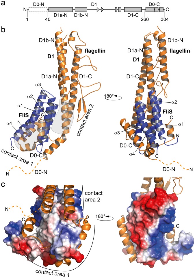
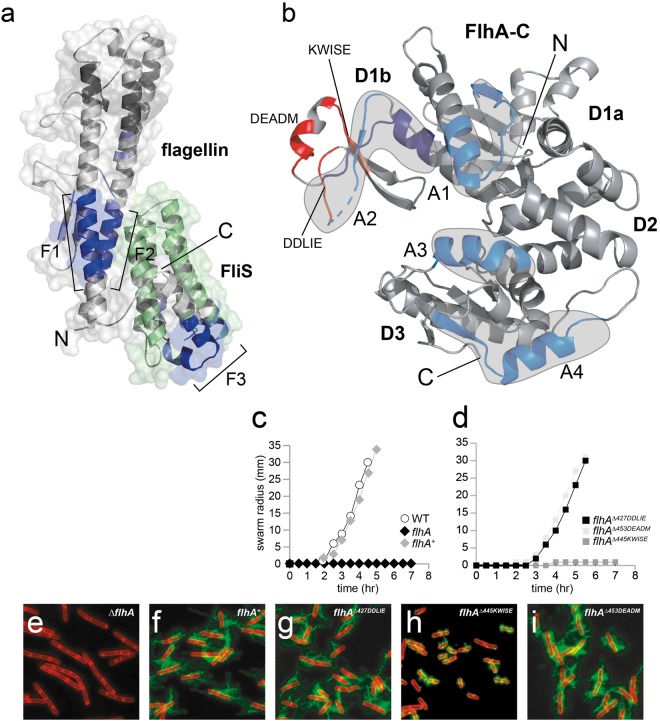
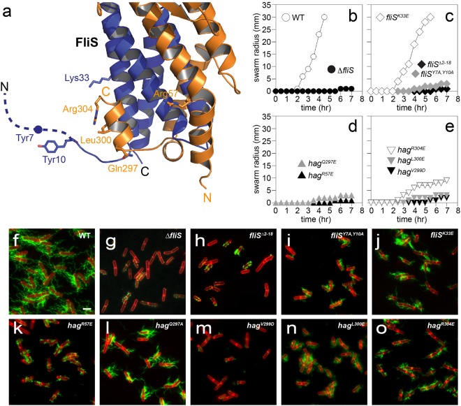
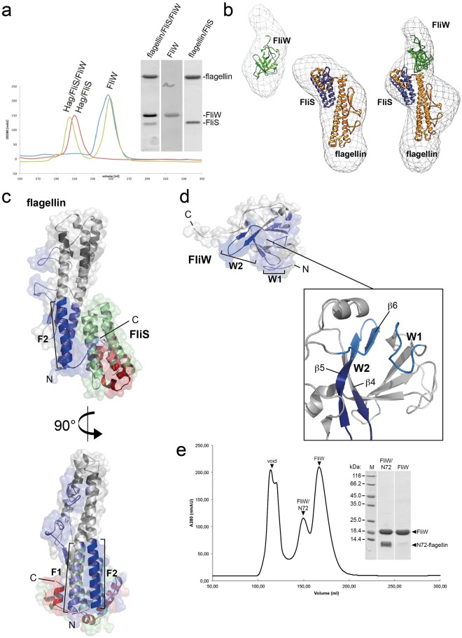
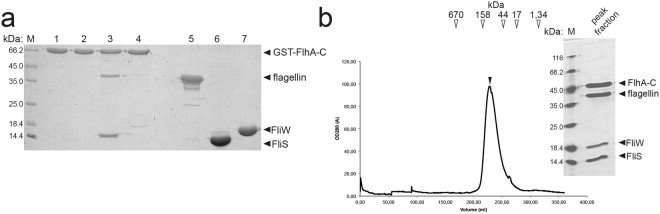
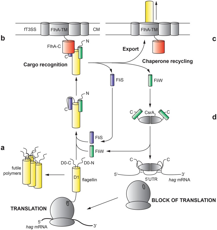
Flagellin is amongst the most abundant proteins in flagellated bacterial species and constitutes the major building block of the flagellar filament. The proteins FliW and FliS serve in the post-transcriptional control of flagellin and guide the protein to the flagellar type III secretion system (fT3SS), respectively. Here, we present the high-resolution structure of FliS/flagellin heterodimer and show that FliS and FliW bind to opposing interfaces located at the N- and C-termini of flagellin. The FliS/flagellin/FliW heterotrimer is able to interact with FlhA-C suggesting that FliW and FliS are released during flagellin export. After release, FliW and FliS are recycled to execute a new round of post-transcriptional regulation and targeting. Taken together, our study provides a mechanism explaining how FliW and FliS synchronize the production of flagellin with the capacity of the fT3SS to secrete flagellin.


Conflict of interest statement

The authors declare no competing interests.

Figures

Figure 1
Figure 1
Crystal structure of the flagellin/FliS heterodimer. (a) Domain organization of flagellin. (b) Crystal structure of flagellin/FliS as a cartoon representation in two orientations. flagellin and FliS are shown in orange and blue, respectively. ‘N’ and ‘C’ indicate N- and C-termini, respectively. The disordered N-terminus of flagellin is shown as dashed line. Contact areas between flagellin and FliS are encircled in grey. (c) Electrostatic surface potential of FliS within the contact area of flagellin to FliS. The flagellin-binding pocket is of hydrophobic and polar nature (left side), whereas the opposite site of FliS is highly charged (right side).
Figure 2
Figure 2
Recruitment of the FliS/flagellin/FliW complex to FlhA-C. (a) HDX analysis of flagellin/FliS in the presence of FlhA-C. Difference in HDX-labelling is shown in different shades of blue (dark blue: strong protection; light blue: weak protection) in Dalton. Regions in the cartoon representation of flagellin/FliS that exchange less in the presence of FlhA-C are highlighted (F1, F2 and F3). FliS is shown in green as no HDX-data were obtained. (b) HDX analysis of FlhA-C in the presence of flagellin/FliS. The difference in HDX-labelling is shown. Blue regions in the cartoon representation of FlhA-C exchange less in the presence of flagellin/FliS and are encircled in grey (A1, A2, A3 and A4). Peptides deleted within the D1b domain are highlighted in red. (c,d) Quantitative swarm expansion assay for flhA mutant and complementation strains: WT is swarming proficient and covers a 0.7% LB agar plate in about 4.5 hours but ΔflhA is non-swarming. The flhAΔ445KWISE mutant strain shows also a strong swarming deficiency (ei) Fluorescence microscopy of B. subtilis showing deficiency in filament assembly of a flhA and flhAΔ445KWISE mutant. Scale bars are 2 µm.
Figure 3
Figure 3
Physiology of flagellin/FliS binding to FlhA-C. (a) Projection of different amino acid exchanges onto the structure of flagellin/FliS. Flagellin is shown in orange, while FliS is depicted in blue. Dashed lines indicate disordered regions. (be) Quantitative swarm expansion assay for fliS and flagellin (hag) mutants: WT is swarming proficient and covers a 0.7% LB agar plate in about 4.5 hours but ΔfliS is non-swarming. Both fliSΔ2–18 and fliSY7A,Y10A phenocopy a ΔfliS mutant for swarming. fliSK33E is swarming proficient. hagR57E confers a non-swarming phenotype while all hag mutants except hagV299D show almost no swarming motility. (fo) Fluorescence microscopy of B. subtilis filaments shows deficiency of ΔfliS, fliSΔ2-18 and fliSY7A,Y10A mutants to secrete flagellin. fliSK33E shows short flagellar filaments. hagR57E, hagV299D, hagL300E and hagR304E show almost no flagellar filaments, while hagQ297A exhibits short flagellar filaments. Scale bars are 2 µm.
Figure 4
Figure 4
SAXS- and HDX-analysis of the FliS/flagellin/FliW interaction. (a) Size-exclusion chromatograms (SEC) of flagellin/FliS (red), FliW (blue) and FliS/flagellin/FliW (green). The inset shows a Coomasse-stained SDS PAGE of the peak fractions. (b) SAXS analysis of FliW, flagellin/FliS and FliS/flagellin/FliW. The crystal structures have been fitted to the SAXS-density with the docking algorithm implemented in Chimera. (c) HDX analysis of flagellin/FliS versus flagellin/FliW. The difference in HDX-labelling is shown from blue (less exchange) to red (more exchange) in Dalton. Blue regions in the cartoon representation of flagellin/FliS exchange less in the presence of FliW (F1 and F2), whereas red regions get unprotected. (d) HDX analysis of FliW versus flagellin/FliW. The difference in HDX-labelling is shown from blue (less exchange) to red (more exchange) in Dalton. Blue regions in the cartoon representation of FliW exchange less in the presence of flagellin (W1 and W2). (e) SEC-chromatogram of a flagellinN72/FliW complex (left) and a Coomassie-stained SDS-PAGE of the two peak fractions (right).
Figure 5
Figure 5
FliW is not a prerequisite for flagellin recognition by FlhA-C. (a) Coomassie-stained SDS-PAGE of a pulldown assay employing GST-tagged FlhA-C. Flagellin alone does not interacts with FlhA-C (lane 2). FliS alone is able to recruit flagellin to FlhA-C (lane 3), while FliW alone is not (lane 4). Lanes 1, 5, 6 and 7 represent the input of purified GST-FlhA-C, Flagellin, FliS and FliW, respectively. (b) Reconstitution of the FliS/flagellin/FliW/FlhA-C complex on size exclusion chromatography shows an apparent heterotetramer. Arrows indicate the protein size standard in kDa. The inset shows a Coomassie-stained SDS-PAGE of the peak fraction.
Figure 6
Figure 6
Model for coupling homeostasis and type-III-secretion of flagellin. The figure depicts how FliS, FliW and CsrA act in concert to couple flagellin homeostasis with its export by the fT3SS. Flagellin is shown in yellow, FliW in green, FliS in blue and FlhA-C in red. CsrA was not part of this study and is shown in grey. (a) FliS and FliW bind to flagellin once it has emerged from the ribosome to prevent formation of futile aggregates. (b) The FliS/flagellin/FliW complex is recognized by the type III secretion system via FlhA-C. (c) FliS and FliW are recycled after flagellin secretion. (d) Free FliW can sequester CsrA from the hag leader transcript, thereby allowing the next round of flagellin translation.

References

    1. Diepold, A. & Armitage, J. P. Type III secretion systems: the bacterial flagellum and the injectisome. Philos. Trans. R. Soc. Lond. B. Biol. Sci. 370 (2015). - PMC - PubMed
    1. Chevance FFV, Hughes KT. Coordinating assembly of a bacterial macromolecular machine. Nat. Rev. Microbiol. 2008;6:455–65. doi: 10.1038/nrmicro1887. - DOI - PMC - PubMed
    1. Mukherjee S, Kearns DB. The structure and regulation of flagella in Bacillus subtilis. Annu. Rev. Genet. 2014;48:319–40. doi: 10.1146/annurev-genet-120213-092406. - DOI - PMC - PubMed
    1. Altegoer F, Bange G. Undiscovered regions on the molecular landscape of flagellar assembly. Curr. Opin. Microbiol. 2015;28:98–105. doi: 10.1016/j.mib.2015.08.011. - DOI - PubMed
    1. Kinoshita M, Hara N, Imada K, Namba K, Minamino T. Interactions of bacterial flagellar chaperone-substrate complexes with FlhA contribute to co-ordinating assembly of the flagellar filament. Mol. Microbiol. 2013;90:1249–61. doi: 10.1111/mmi.12430. - DOI - PubMed

Publication types

MeSH terms

Substances

LinkOut - more resources