. 2018 Aug 1;9(8):828.
doi: 10.1038/s41419-018-0892-3.

Double-edged sword of gonadotropin-releasing hormone (GnRH): A novel role of GnRH in the multiple beneficial functions of endometrial stem cells

Affiliations

Double-edged sword of gonadotropin-releasing hormone (GnRH): A novel role of GnRH in the multiple beneficial functions of endometrial stem cells

Se-Ra Park et al. Cell Death Dis. .

Abstract

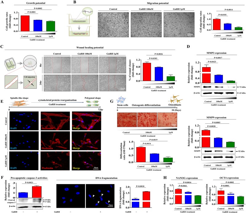
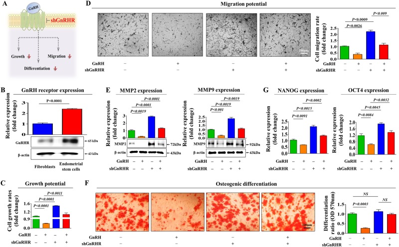
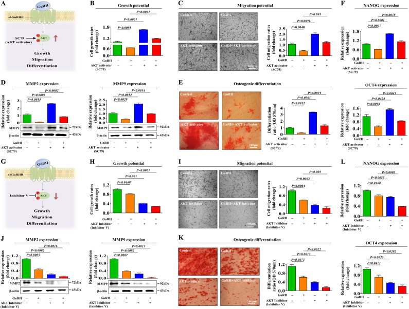
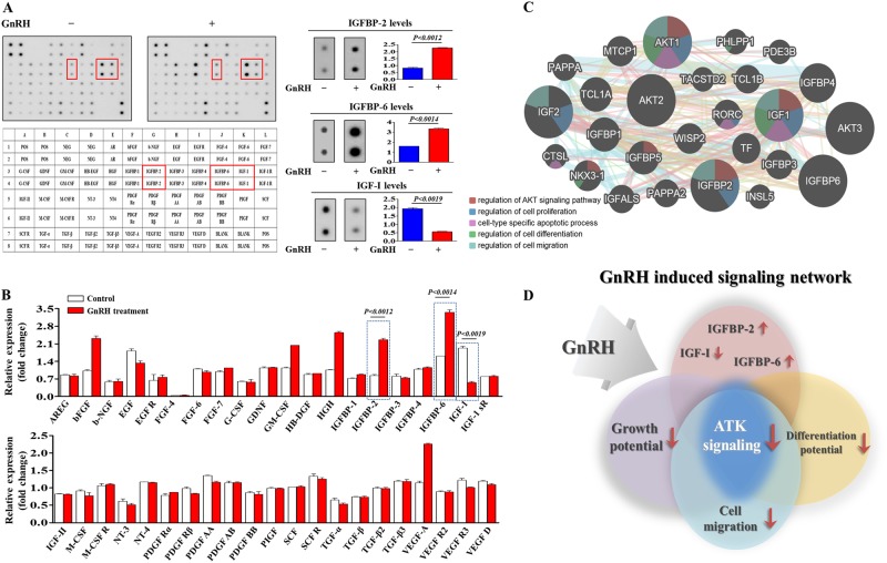
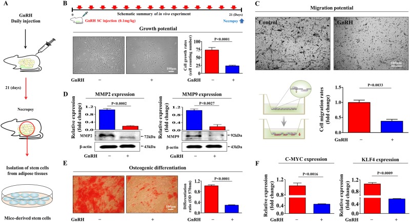
Gonadotropin-releasing hormone (GnRH) stimulates the synthesis and release of gonadotropins, which induce estrogen production and subsequent ovulation. Therefore, long-term GnRH exposure to regulate ovarian hyperstimulation is recognized as the gold standard for most in vitro fertilization (IVF) strategies. However, one of the most disappointing aspects of current IVF technology is relatively low rate (between 35 and 50%) of positive pregnancy outcomes, and the major reason for this high cancellation rate has not yet been revealed. Previous studies have demonstrated that resident stem cell deficiency limits the cyclic regenerative capacity of the endometrium and subsequently increases pregnancy failure rates. Therefore, we hypothesized that long-term GnRH exposure directly damages endometrial stem cells and consequently negatively affects pregnancy outcomes in GnRH-based IVF. In addition to their well-known roles in regulating the hypothalamus-pituitary-gonadal axis, GnRH and its receptors also localize in the extra-hypothalamic endometrium, suggesting a possible non-canonical role in endometrial stem cells. Consistent with our hypothesis, we show for the first time that GnRH suppresses the multiple beneficial functions of endometrial stem cells via the PI3K/Akt signaling pathway in vitro and in vivo. To the best of our knowledge, this is the first study to focus on the direct effects of GnRH on the regenerative potential of stem cells, and the findings will facilitate the development of more promising IVF strategies.


Conflict of interest statement

The authors declare that they have no conflict of interest.

Figures

Fig. 1
Fig. 1. GnRH suppresses multiple beneficial functions of endometrial stem cells in vitro.
The inhibition of endometrial stem cell viability by 1 µM GnRH at 72 h was determined by an MTT assay. Stem cell viability (%) was calculated as a percent of the vehicle control (a). Endometrial stem cells were treated with GnRH for 72 h, and the effect of GnRH on stem cell migration ability was then evaluated using the transwell migration assay. GnRH treatment significantly decreased stem cell migration across the membrane compared with the negative control (b). The effects of GnRH on stem cell migration were further evaluated using a scratch assay. The migration of stem cells treated with GnRH was slower than that of cells treated with vehicle. (c). The relative expression levels of key positive regulators of cell migration (MMP-2/9) were assessed using western blotting (d). GnRH-induced actin filament disorganization and morphological changes in stem cells were visualized by staining actin filaments with phalloidin (e). Elevated levels of cleaved caspase-3 following GnRH treatment were assessed by western blotting. GnRH-induced apoptotic DNA fragmentation and condensation were visualized using DAPI staining (f). Confluent stem cells were cultured in osteogenic medium with or without GnRH. The effects of GnRH on osteoblast differentiation were determined by alizarin red staining. The relative quantification of calcium mineral content was performed by measuring the absorbance at 570 nm (g). Real-time PCR results showed the changes in the expression of the stem cell markers NANOG and OCT4 after GnRH treatment for 72 h (h). DAPI staining was used to label the nuclei. β-actin was used as the internal control. The data are presented as the mean ± SD of three independent experiments
Fig. 2
Fig. 2. GnRH inhibits multiple beneficial stem cell functions through its cognate receptor.
Schematic representation of the experimental protocol as described in the materials and methods section (a). Fibroblasts and endometrial stem cells were incubated under standard culture conditions, and the expression levels of GnRH-R were assessed by western blotting (b). Endometrial stem cells were treated with 1 µM GnRH alone or in combination with shRNA targeting GnRH-R; subsequent changes in cell viability were measured by an MTT assay (c). Changes in migratory capacity were measured by the transwell assay (d) and western blotting for MMP-2 and MMP-9 (e). The ability of GnRH-R knockdown to attenuate the GnRH-induced suppression of osteoblast differentiation was determined by alizarin red staining. The relative quantification of calcium mineral content was performed by measuring the absorbance at 570 nm (f). Real-time PCR results showed the changes in the expression of the stem cell markers NANOG and OCT4 after GnRH treatment for 72 h alone or in combination with GnRH-R shRNA (g). β-actin was used as the internal control. The data are presented as the mean ± SD of three independent experiments
Fig. 3
Fig. 3. The inhibitory effects of GnRH on PI3K/Akt signaling in vitro and in vivo.
Schematic representation of the experimental protocol as described in the materials and methods section (a). Endometrial stem cells were treated for 10 min with or without GnRH (1 µM). The cells were then lysed, and the protein contents were analyzed by western blotting using antibodies targeting the phosphorylated forms of PI3K and Akt. The phosphorylation levels of these signaling molecules were significantly decreased in stem cells treated with GnRH (b). Endometrial stem cells were treated with 1 µM GnRH alone or in combination with shRNA targeting GnRH-R; subsequent changes in the phosphorylation levels of PI3K and Akt were measured via western blotting (c). Mice were treated daily for 21 days with GnRH (0.1 mg/kg, subcutaneously) or vehicle (PBS). Stem cells were isolated from mouse adipose tissues, and changes in the phosphorylation levels of PI3K and Akt were measured via western blotting (d). β-actin was used as the internal control. The data are presented as the mean ± SD of three independent experiments
Fig. 4
Fig. 4. Activation or inhibition of Akt signaling attenuates the GnRH-induced effects on multiple stem cell functions.
Schematic representation of the experimental protocol as described in the materials and methods section (a). Endometrial stem cells were pretreated with the Akt activator SC79 (10 µM) for 24 h prior to treatment with 1 µM GnRH for 48 h, and the changes in cell viability were determined by an MTT assay. Stem cell viability (%) was calculated as a percent of the vehicle control (b). The changes in migratory capacity were measured via the transwell assay (c) and western blotting for MMP-2 and MMP-9 (d). The changes in osteoblast differentiation were determined by alizarin red staining. The relative quantification of calcium mineral content was performed by measuring the absorbance at 570 nm (e). The ability of SC79 to attenuate the GnRH-induced inhibition of stem cell marker expression (NANOG and OCT4) was determined by real-time PCR (f). Schematic representation of the experimental protocol as described in the materials and methods section (g). Endometrial stem cells were pretreated with Akt inhibitor V (10 µM) for 24 h prior to treatment with 1 µM GnRH for 48 h, and the changes in cell viability were determined by an MTT assay. Stem cell viability (%) was calculated as a percent of the vehicle control (h). The changes in migratory capacity were measured via the transwell assay (i) and western blotting for MMP-2 and MMP-9 (j). The changes in osteoblast differentiation were determined by alizarin red staining. The relative quantification of calcium mineral content was performed by measuring the absorbance at 570 nm (k). The synergism between Akt inhibitor V and GnRH in inhibiting stem cell marker expression (NANOG and OCT4) was determined by real-time PCR (l). β-actin was used as the internal control. The data are presented as the mean ± SD of three independent experiments
Fig. 5
Fig. 5. GnRH-induced secreted proteins are associated with PI3K/Akt signaling.
Human growth factor antibody array analysis was performed using GnRH-treated and control samples. The membrane was printed with antibodies for 40 growth factors, cytokines, and receptors, with four positive and four negative controls in the upper and lower left corners. Three growth factors or related proteins (IGF-I, IGFBP-2, and IGFBP-6) were markedly enriched in the GnRH-treated groups compared with the control groups (a, b). Signaling network analysis was performed using GeneMANIA (http://www.genemania.org) to predict the connections between the three growth factors and PI3K/Akt signaling. The results revealed a positive relationship between each of the three factors (IGF-I, IGFBP-2, and IGFBP-6) and PI3K/Akt signaling (c, d). The data are presented as the mean ± SD of three independent experiments
Fig. 6
Fig. 6. GnRH inhibits multiple beneficial functions of stem cells in vivo.
Schematic representation of the experimental protocol as described in the materials and methods section (a). Mice were treated daily for 21 days with GnRH (0.1 mg/kg, intraperitoneally) or vehicle (PBS). Stem cells were isolated from mouse adipose tissue, and changes in cell viability were determined by an MTT assay. Stem cell viability (%) was calculated as a percent of the vehicle control (b). The changes in migratory capacity were measured via the transwell assay (c) and western blotting for MMP-2 and MMP-9 (d). The changes in osteoblast differentiation were determined by alizarin red staining. The relative quantification of calcium mineral content was performed by measuring the absorbance at 570 nm (e). Real-time PCR results showed the changes in the expression of the mouse stem cell markers C-MYC and KLF4 after GnRH treatment in vivo (f). β-actin was used as the internal control. The data are presented as the mean ± SD of three independent experiments

References

    1. Limonta P, Moretti RM, Marelli MM, Motta M. The biology of gonadotropin hormone-releasing hormone: role in the control of tumor growth and progression in humans. Front. Neuroendocrinol. 2003;24:279–295. doi: 10.1016/j.yfrne.2003.10.003. - DOI - PubMed
    1. Neill JD. GnRH and GnRH receptor genes in the human genome. Endocrinology. 2002;143:737–743. doi: 10.1210/endo.143.3.8705. - DOI - PubMed
    1. Kumar P, Sharma A. Gonadotropin-releasing hormone analogs: Understanding advantages and limitations. J. Hum. Reprod. Sci. 2014;7:170–174. doi: 10.4103/0974-1208.142476. - DOI - PMC - PubMed
    1. Depalo R, et al. GnRH agonist versus GnRH antagonist in in vitro fertilization and embryo transfer (IVF/ET) Reprod. Biol. Endocrinol. 2012;10:26. doi: 10.1186/1477-7827-10-26. - DOI - PMC - PubMed
    1. Lai Q, et al. Comparison of the GnRH agonist and antagonist protocol on the same patients in assisted reproduction during controlled ovarian stimulation cycles. Int. J. Clin. Exp. Pathol. 2013;6:1903–1910. - PMC - PubMed

Publication types

MeSH terms

Substances

LinkOut - more resources