. 2018 Aug 1;9(1):3020.
doi: 10.1038/s41467-018-05412-0.

Regulation of T cell afferent lymphatic migration by targeting LTβR-mediated non-classical NFκB signaling

Affiliations

Regulation of T cell afferent lymphatic migration by targeting LTβR-mediated non-classical NFκB signaling

Wenji Piao et al. Nat Commun. .

Erratum in

Abstract

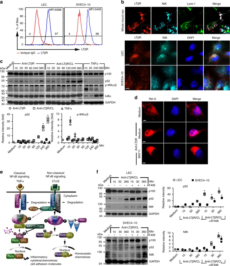
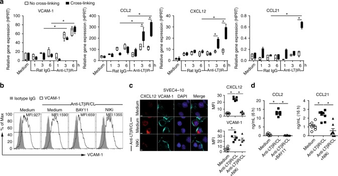
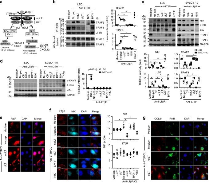
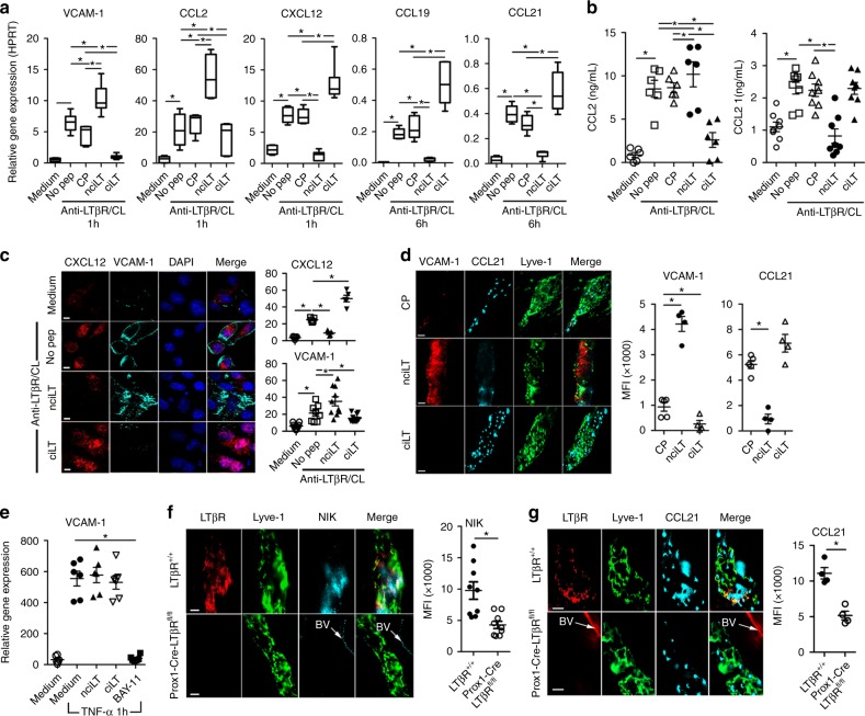
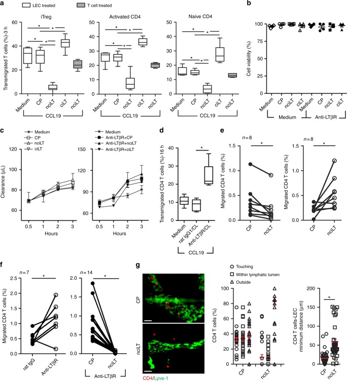
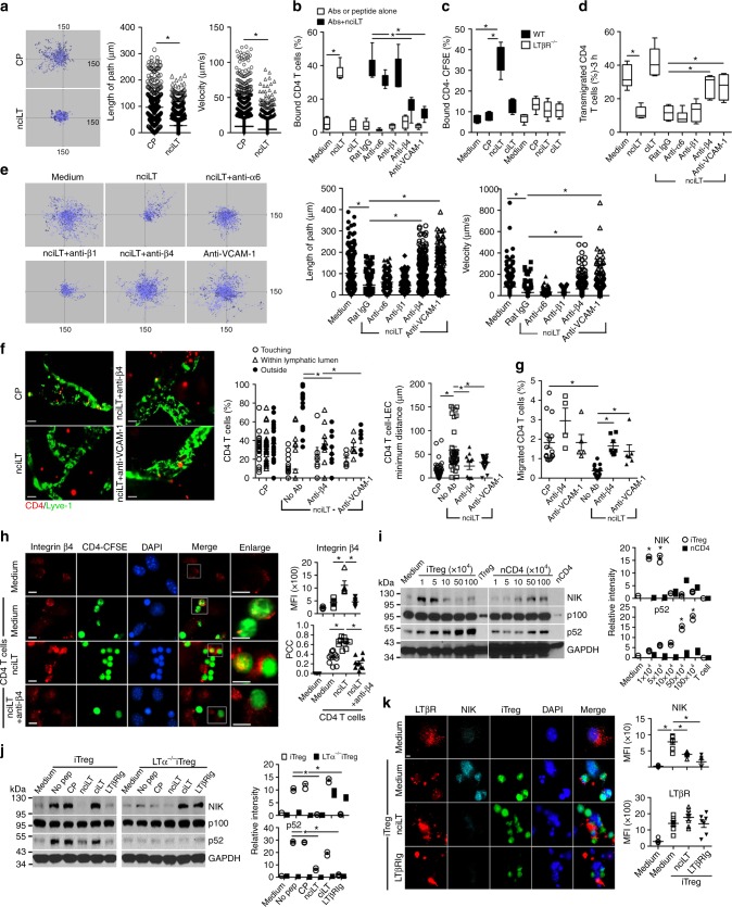
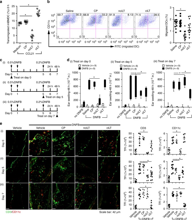
Lymphotoxin-beta receptor (LTβR) signaling in lymphatic endothelial cells (LEC) regulates leukocyte afferent lymphatic transendothelial migration (TEM). The function of individual signaling pathways for different leukocyte subsets is currently unknown. Here, we show that LTβR signals predominantly via the constitutive and ligand-driven non-classical NIK pathway. Targeting LTβR-NIK by an LTβR-derived decoy peptide (nciLT) suppresses the production of chemokines CCL21 and CXCL12, and enhances the expression of classical NFκB-driven VCAM-1 and integrin β4 to retain T cells on LEC and precludes T cell and dendritic cell TEM. nciLT inhibits contact hypersensitivity (CHS) at both the sensitization and elicitation stages, likely by inhibiting leukocyte migration. By contrast, targeting LTβR-classical NFκB signaling during the elicitation and resolution stages attenuates CHS, possibly by promoting leukocyte egress. These findings demonstrate the importance of LTβR signaling in leukocyte migration and LEC and lymphatic vessel function, and show that antagonist peptides may serve as lead compounds for therapeutic applications.


Conflict of interest statement

The authors declare no competing interests.

Figures

Fig. 1
Fig. 1
Preferential non-classical NFκB signaling induced by LTβR activation in LEC. a Flow cytometry analysis of LTβR expression on murine primary LEC and SVEC4-10. MFI mean fluorescence intensity. b Immunohistochemistry of LTβR and NIK expression on mouse whole-mount ear, primary LEC, and SVEC4-10. Magnification ×20, ×60 (inset); scale bar 10 μm. c Mouse primary LEC stimulated with 2 μg/mL 3C8 anti-LTβR mAb or 20 ng/mL TNFα for the indicated times. For crosslinking (CL), cells incubated with 2 μg/mL anti-LTβR mAb at 4 °C, washed, and then crosslinked with 2 μg/mL mouse anti-rat IgG1 for the indicated times. Cell lysates immunoblotted for p100, p52, phosphorylated IKKα/β, IκBα, and GAPDH. The bar graphs represent the relative band intensities (mean ± SEM) from three independent experiments. *p< 0.05 by one-way ANOVA. d Nuclear translocation of RelA/p65 in LEC stimulated with 2 μg/mL anti-LTβR for 10 min. Magnification ×60; scale bar 4 μm. e Diagram of TNFα and LTβR-mediated classical and non-classical NFκB signaling. f Mouse primary LEC or SVEC4-10 treated as in c, along with 25 μM AT406. Cell lysates immunoblotted for p100, p52, NIK, and GAPDH. The bar graphs represent the relative band intensities (mean ± SEM) from three independent experiments. *p< 0.05 by one-way ANOVA
Fig. 2
Fig. 2
Agonistic anti-LTβR mAb induces inflammatory and homeostatic chemokines and cell adhesion molecules in LEC. a qRT-PCR of indicated genes induced by agonistic anti-LTβR mAb or isotype rat IgG crosslinked with or without mouse anti-rat IgG1 in LEC at the indicated times. Treated as in Fig. 1.b Flow cytometry analysis of VCAM-1 expression on LEC after treatment with anti-LTβR mAb plus crosslinking, with or without 25 μM NFκB inhibitor BAY11-7085 or 50 μM NIKi for 3 h. c LTβR-induced CXCL12 and VCAM-1 expression on SVEC4-10 treated with anti-LTβR mAb plus crosslinking, with or without NIKi (50 μM) for 3 h. Magnification ×60; scale bar 5 μm. d Secretion of CCL2 and CCL21 by LEC stimulated with anti-LTβR mAb plus crosslinking, with or without BAY11-7085 (25 μM) or NIKi (50 μM) measured at the indicated times. *p< 0.05 by one-way ANOVA (a, d) or by Student’s t-test (c)
Fig. 3
Fig. 3
Targeting of LTβR-mediated classical and non-classical NFκB signaling pathways by LTβR-specific peptides. a Diagram of peptide selective blockade of separate arms of LTβR signaling. b Immunoprecipitation of LTβR complex with anti-LTβR in lysates of LEC pretreated with the indicated peptides (20 μM) and then stimulated with anti-LTβR mAb (2 μg/mL) for 10 min. Complexes run on SDS-PAGE, and immune blotted with anti-TRAF2, anti-TRAF3, and anti-LTβR. c, d LEC and SVEC4-10 pretreated with indicated peptides (20 μM) or inhibitors (25 μM BAY11-7085; 50 μM NIKi) and then stimulated with anti-LTβR (2 μg/mL) for 6 h (c) or 10 min (d). In panel d, SVEC4-10 stimulated with 20 ng/mL TNFa. Cell lysates immune blotted for p100, p52, NIK, TRAF2, and TRAF3 (c); for IKKα/β, and for IκBα phosphorylation and degradation (d). e Cells treated as in (d); immunohistochemistry of RelA. Magnification ×60; scale bar 4 μm. f, g Cells treated as in (c). Immunohistochemistry of LTβR and NIK in SVEC4-10 (f); CCL21 and RelB in LEC (g). Magnification ×60; scale bar 8 μm (f) or 4 μm (g). The bar graphs in (bd) represent the relative band intensities (mean ± SEM) from three independent experiments. *p< 0.05 by one-way ANOVA
Fig. 4
Fig. 4
Regulation of gene profiles by the LTβR NFκB-blocking peptides. a qRT-PCR of VCAM-1 and chemokines in LEC stimulated with anti-LTβR mAb (2 μg/mL) plus CL with mouse anti-rat IgG1 (2 μg/mL) for the indicated times. b ELISA for CCL2 and CCL21 in the LEC supernatants after 4 or 16 h stimulation as in a, respectively. c CXCL12 and VCAM-1 expression in LEC after 3 h stimulation as in a. Magnification ×40; scale bar 20 μm. d C57BL/6 mice injected with 5 nmol/ear control peptide (CP), nciLT or ciLT; after 3 h ears were fixed for whole-mount ear staining. Magnification ×20; scale bar 50 μm. e qRT-PCR of VCAM-1 in LEC stimulated with TNFα (20 ng/mL) for 1 h. f, g Whole-mount ear staining of NIK and CCL21 in Prox1-Cre-ERT2+/−LTβRfl/fl mice 10 days after tamoxifen treatment (0.125 mg/g i.p. for 5 consecutive days). BV blood vessel. Magnification ×60; scale bar 10 μm. *p < 0.05 by one-way ANOVA (ae) or by Student’s t-test (f, g)
Fig. 5
Fig. 5
LTβR peptides that block non-classical NFκB signaling pathway inhibit CD4 T cell migration across LEC. a LEC cell layers pretreated with the indicated peptides (20 μM) for 30 min at 37 °C and then Foxp3+CD4+CD25+ iTregs, Foxp3CD25CD4+aCD4, or naïve CD4 cells loaded into upper chamber and migrated toward CCL19 (50 ng/mL) for 3 h. b LEC incubated with 20 μM nciLT or ciLT peptides for 3 h without or with stimulation with 2 μg/mL anti-LTβR mAb. Cell viability determined by MTT incorporation assay. c Time-course monitoring of 0.75% albumin/Evans Blue diffusion across LEC cell layer in Boyden chamber treated as in b. d LEC cell layers pretreated with anti-LTβR mAb or isotype rat IgG for 30 min at 4 °C, washed and CL with mouse anti-rat IgG1 for 30 min at 37 °C, followed by 16 h incubation, then naïve CD4 cells loaded into the upper chambers and migrated toward CCL19 (50 ng/mL) for 3 h. e, f Footpad-popliteal LN migration assay. 1 × 106 CFSE-labelled naïve CD4 T cells mixed with 5 nmol CP (left side), 5 nmol nciLT (right side), or 5 nmol ciLT (right side) without (e) or with 1 μg anti-LTβR mAb (f), injected into hind footpads. After 16 h popliteal LNs collected and analyzed with flow cytometry. g Ear whole-mount staining and migration. CFSE-naïve CD4 T cells injected into ear pinnae pretreated with peptides (5 nmol/ear), and collected after 16 h post injection. Images of T cells and Lyve-1+ lymphatic vessels (left). Magnification ×20; scale bar 50 μm. Position (center) and distance (right) of T cells with respect to lymphatic vessels. ag Mean ± SEM of at least three independent experiments. *p < 0.05 by one-way ANOVA (ad) or by Student’s t-test (eg)
Fig. 6
Fig. 6
Inhibition of T cell stimulated LTαβ–LTβR NIK pathway on LEC enhances T cell binding to LEC through integrin β4 and VCAM-1. a Time-lapse, 2-D microscopy of CD4 T cell migration across LEC. LEC layers in Boyden chambers treated with 20 μM CP or nciLT for 30 min at 37 °C, washed and naïve CD4-CFSE T cell migration to CCL19 (50 ng/mL) monitored for 3 h by live imaging. b Binding of naïve CD4 T cells to LEC pretreated with the indicated peptides (20 μM) with or without anti-integrin mAbs (2 μg/mL) or anti-VCAM-1 mAb (3 μg/mL) for 30 min at 37 °C. c Binding of naïve CD4 T cells to wild type (WT) or LTβR−/− LEC pretreated with the indicated peptides (20 μM). d LEC layers in Boyden chamber treated as in b, then naïve CD4 T cells migrated across LEC. e LEC layers treated with peptides and mAbs as in b for 30 min at 37 °C, washed, and naïve CD4 T cell migration toward CCL19 (50 ng/mL) monitored for 3 h of live imaging. f Ear whole-mount staining and migration. Pinnae injected with nciLT (5 nmol/ear) with or without anti-integrin β4 (5 μg) or anti-VCAM-1 mAbs (15 μg) 30 min before transfer of naïve CSFE-labeled CD4 T cells (1 × 106/ear). After 16 h, ears collected and stained for Lyve-1. Images of T cells and Lyve-1+ lymphatic vessels (left). Magnification ×20; scale bar 50 μm. Position (center) and distance (right) of T cells with respect to lymphatic vessels. g Footpad-popliteal LN migration assay. Hind footpads injected with the same doses of peptide or antibodies as in f, then 1 × 106 naïve CD4-CFSE T cells transferred. After 16 h, popliteal LN collected and analyzed with flow cytometry. h Expression of cell surface integrins on primary LEC. LEC treated with 20 μM nciLT with or without anti-integrin β4 mAb (2 μg/mL) prior to co-culture with 2 × 105 naïve CD4-CFSE for 3 h. Unbound T cells gently washed away before fixation. PCC Pearson’s correlation coefficient. Magnification ×60; scale bar 5 μm. i, j Immunoblot of NIK activation in LEC pretreated with the indicated peptides (20 μM) or LTβRIg (2 μg/mL) for 30 min, then incubated with various numbers of purified iTreg or naïve CD4 T cells (i) or 5 × 105 wild type or LTα-deficient iTreg (j) for 6 h. The bar graphs represent the relative band intensities (mean ± SEM) from three independent experiments. k Expression of LTβR and NIK in primary LEC pretreated with 20 μM nciLT or 2 mg/mL LTβRIg for 30 min at 37 °C, and co-cultured with 2 × 105 iTreg-CFSE for 6 h. Magnification ×60; scale bar 5 μm. *p < 0.05 by Students’ t-test (a) or by one-way ANOVA (bk)
Fig. 7
Fig. 7
Inhibition of LTβR signaling alters CHS and leukocyte migration. a In vitro DC migration to CCL21 (50 ng/mL) through LEC treated with the indicated peptides (20 μM). b In vivo DC migration assay. i.d. injection of the indicated peptides (20 nmol) to the shaved abdomen, followed 30 min later by FITC painting. After 16 h, inguinal and axillary LNs collected and analyzed with flow cytometry. c Experimental design schematic. 20 nmol of indicated peptides injected i.d. in the abdomen (100 μL, day 0) or 10 nmol in each ear (30 μL, day 5 or day 7) 30 min before DNFB painting on days 0 or 5. d, e CHS analyzed by ear swelling measured 48 h after DNFB challenge (d), or immunohistochemistry for CD11c+ DC and CD3+ T cells (e). Magnification ×20; scale bar 42 μm. TFI total fluorescence intensity. Representative of three independent experiments with 3 mice/group. *p < 0.05 by one-way ANOVA

References

    1. Butcher EC, Picker LJ. Lymphocyte homing and homeostasis. Science. 1996;272:60–66. doi: 10.1126/science.272.5258.60. - DOI - PubMed
    1. Schwab SR, Cyster JG. Finding a way out: lymphocyte egress from lymphoid organs. Nat. Immunol. 2007;8:1295–1301. doi: 10.1038/ni1545. - DOI - PubMed
    1. Brinkman CC, et al. Treg engage lymphotoxin beta receptor for afferent lymphatic transendothelial migration. Nat. Commun. 2016;7:12021. doi: 10.1038/ncomms12021. - DOI - PMC - PubMed
    1. Zhang N, et al. Regulatory T cells sequentially migrate from inflamed tissues to draining lymph nodes to suppress the alloimmune response. Immunity. 2009;30:458–469. doi: 10.1016/j.immuni.2008.12.022. - DOI - PMC - PubMed
    1. Xiong Y, Ahmad S, Iwami D, Brinkman CC, Bromberg JS. T-bet regulates natural regulatory T cell afferent lymphatic migration and suppressive function. J. Immunol. 2016;196:2526–2540. doi: 10.4049/jimmunol.1502537. - DOI - PMC - PubMed

Publication types

MeSH terms