. 2018 Aug;560(7719):509-512.
doi: 10.1038/s41586-018-0396-4. Epub 2018 Aug 1.

Inositol phosphates are assembly co-factors for HIV-1

Affiliations

Inositol phosphates are assembly co-factors for HIV-1

Robert A Dick et al. Nature. 2018 Aug.

Erratum in

Abstract

A short, 14-amino-acid segment called SP1, located in the Gag structural protein1, has a critical role during the formation of the HIV-1 virus particle. During virus assembly, the SP1 peptide and seven preceding residues fold into a six-helix bundle, which holds together the Gag hexamer and facilitates the formation of a curved immature hexagonal lattice underneath the viral membrane2,3. Upon completion of assembly and budding, proteolytic cleavage of Gag leads to virus maturation, in which the immature lattice is broken down; the liberated CA domain of Gag then re-assembles into the mature conical capsid that encloses the viral genome and associated enzymes. Folding and proteolysis of the six-helix bundle are crucial rate-limiting steps of both Gag assembly and disassembly, and the six-helix bundle is an established target of HIV-1 inhibitors4,5. Here, using a combination of structural and functional analyses, we show that inositol hexakisphosphate (InsP6, also known as IP6) facilitates the formation of the six-helix bundle and assembly of the immature HIV-1 Gag lattice. IP6 makes ionic contacts with two rings of lysine residues at the centre of the Gag hexamer. Proteolytic cleavage then unmasks an alternative binding site, where IP6 interaction promotes the assembly of the mature capsid lattice. These studies identify IP6 as a naturally occurring small molecule that promotes both assembly and maturation of HIV-1.


Figures

Extended Data Fig. 1 ∣
Extended Data Fig. 1 ∣. Effect of acidic molecules on immature s-CANC assembly.
a, Representative negative stain EM images. Scale bars, 200 nm. The experiment was repeated two times with similar results. b, Number of immature VLPs per 55 µm2. n=5 and mean above box plots; center lines show the medians; box limits indicate the 25th and 75th percentiles as determined by R software; whiskers extend to minimum and maximum values.
Extended Data Fig. 2 ∣
Extended Data Fig. 2 ∣. s-CANC and s-CASP1 VLPs.
a-c, Representative negative stain EM images of s-CANC (a), s-CASP1 (b), and s-CA (c) proteins assembled in the absence of GT25 and in the presence of the indicated IP6 concentrations. Scale bars, 200 nm. d, Diameters of immature VLPs; mean diameter above plot; n below plot. Center lines show the medians; box limits indicate the 25th and 75th percentiles as determined by R software; whiskers extend to minimum and maximum values.
Extended Data Fig. 3 ∣
Extended Data Fig. 3 ∣. Comparison of the HIV-1 Gag cryoEM structure with the CACTDSP1/IP6 crystal structure.
a, The crystal structure of CACTDSP1 bound to IP6 (cyan) was superimposed on a previously described model of the CA-SP1 segment build into cryoEM densities of immature HIV-1 particles (PDB 5L93, orange2). Note the close correspondence in K359 rotamers, which were modeled independently in the two structures. For visualization purposes, only one of the six possible IP6 conformations is displayed. b, RMSD calculations of the crystal structure and PDB 5L93. For full-length (residues 149–237) and CA-SP1 (residues 223–237), the RMSDs were calculated only for the atoms that were modeled in both maps. In case a side chain was not modeled, the entire residue was omitted from the calculation. The overall agreement of the models is very high, indicating that the crystal structure corresponds well with conformations found in the virus. c, The CACTDSP1 bound to IP6 (orange and red, respectively) was fitted into two previously published cryoEM densities from VLPs collected from cells (EMD-2706 and EMD-4017). Both EM maps are shown at 8.8 Å, which is the resolution of the lower resolved map, EMD-2706. In the zoomed insets only the density corresponding to IP6 is shown. Matching of models and maps, and RMSD calculations were performed in Chimera.
Extended Data Fig. 4 ∣
Extended Data Fig. 4 ∣. Interpretation of the IP6 density in the immature CACTDSP1 hexamer structure.
a, Top and side views of the unbiased mFo-DFc difference density (blue mesh, 2σ) ascribed to the bound IP6. Shown are six IP6 molecules docked in six rotationally equivalent positions, consistent with the six-fold rotational symmetric density. b, Top view of the docked IP6 molecules within the CACTDSP1 hexamer. Unbiased mFo-DFc difference densities (blue mesh) are also shown for both the bound IP6 and sidechains of Lys290 (green) and Lys359 (cyan). Density for Lys359 is more pronounced, which we interpret to mean that this residue adopts a more restricted range of rotamers for binding IP6.
Extended Data Fig. 5 ∣
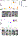
Extended Data Fig. 5 ∣. Quantification of wild type and mutant HIV s-CANC assembly at pH 6 and 8.
a,c, Number of immature (purple) and mature (orange) VLPs per 55 μm2 without (−) and with (+) 10 μM IP6 at pH 6 and pH 8. Mean above and n below box plots. Center lines show the medians; box limits indicate the 25th and 75th percentiles as determined by R software; whiskers extend to minimum and maximum values. b,d, Representative negative stain EM images of WT and mutant s-CANC assembly in the absence (−) and presence (+) of 10 μM IP6 at pH 6 and 8. Scale bar, 400 nm. Repeated three times with similar results. e, Infectivity relative to WT virus of IP6 binding residues mutated to alanine; CA residue numbering in parenthesis. Error bars represent standard deviation, individual data points represented as dots, n from four independent experiments.
Extended Data Fig. 6 ∣
Extended Data Fig. 6 ∣. IP6 modulates the stability of the 6HB.
a, Structural changes observed after 2 μs of molecular dynamics simulations of CACTDSP1 with and without bound IP6. b, RMSDs of the ligand-bound and unbound forms of CACTDSP1 hexamers. c, RMSFs of the central hexamer during the simulation. The RMSF was averaged over the six central monomers; dashed line shows the standard deviation for each residue.
Extended Data Fig. 7 ∣
Extended Data Fig. 7 ∣. Quantification of mature HIV-1 CA assembly and VLP diameter at pH 6.
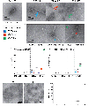
a, Example of CA assembly in the absence of IP6 or mellitic acid. b-c, Representative negative stain EM images of assemblies induced by IP6 (b) and mellitic acid (c). Scale bars, 200 nm. Tubes (T), cones (C), and other (O) morphologies are marked by colored arrowheads. (a, b, c) Repeated four times with similar results. d, Number of CA assembled tubes (blue), cones (orange), and other (green) per 55 μm2 at increasing IP6 concentrations. Mean above and n=5 e, Number of CA assembled tubes (blue), cones (orange), and other (green) per 55 μm2 at increasing mellitic acid concentrations. Mean above and below box plots. f, Representative images of mature VLPs assembled with IP5 and IP6 at 50 mM NaCl. Scale bars, 100 nm. Repeated three times with similar results. g, Number of CA VLPs per 10 µm2 without and with IP3, IP4, IP5, and IP6. Mean above and n=5. (d-e, g) Center lines show the medians; box limits indicate the 25th and 75th percentiles as determined by R software; whiskers extend to minimum and maximum values.
Extended Data Fig. 8 ∣
Extended Data Fig. 8 ∣. Crystal structure of IP6 bound to the mature CA hexamer.
a,b, Top view (a) and side view (b) of a second CA hexamer crystal structure (P212121 space group) showing the protein in yellow ribbons and unbiased mFo-DFc difference density in blue mesh, contoured at 2.5σ. c, Close-up view showing IP6 densities both above and below the ring of Arg18 residues (magenta).
Fig. 1 ∣
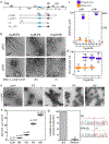
Fig. 1 ∣. IP6 induces assembly of HIV-1 Gag in vitro.
a, Map of the HIV-1 Gag protein, indicating the MA, CA, NC, and p6 domains, and spacer peptides SP1 and SP2. Gag-derived constructs used in this study are shown underneath. Blue bar, major homology region (MHR); purple bar, SP1 helix; NTD and CTD, N-terminal and C-terminal domains of CA; R18, K290, and K359, locations of mutations; N372, C-terminal residue of the s-CASP1 and CACTDSP1 constructs. b, Negative stain EM images of mature and immature VLPs formed by s-CANC (50 μM) at pH 6 and pH 8 in the absence or presence of the indicated molar ratios of IP6 (0–10 μM). Scale bars, 100 nm. c, Number of VLPs per 55/µm2 without (−) and with (+) 10µM IP6 at pH 6 and pH 8; n below and mean above box plots. The experiment was repeated three times with similar results. d, Diameters of immature and mature VLPs; n below and mean above box plots. e, Representative images of s-CANC VLPs assembled at pH 8 in the absence and presence of IP3, IP4, IP5, and IP6. Scale bars, 100 nm. The experiment was repeated two times with similar results. f, Number of VLPs per 55/µm2 without and with 10 µM IP3-IP6; n=5 and mean above box plots g, Parallel transfections of 293FT wildtype (WT) and IPPK KO were performed with a VSV-G-pseudotyped HIV-1 provirus containing GFP, and infectivity was measured on WT 293T cells. Graphs show average and standard deviations of 4 independent experiments; dots show individual data points. Right panels show sequences of total PCR products of the guide RNA target sites from WT and KO cells; guide RNA sequence is underlined in red. (c, d, f). Center lines show the medians; box limits indicate the 25th and 75th percentiles as determined by R software; whiskers extend to minimum and maximum values.
Fig. 2 ∣
Fig. 2 ∣. IP6 interacts with Lys290 and Lys359 in the immature HIV-1 Gag hexamer.
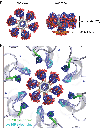
a, IP6-induced assembly of s-CASP1 into immature VLPs. The experiment was repeated four times with similar results. b, IP6-induced assembly of CACTDSP1 into flat micro-crystals. The experiment was repeated six times with similar results. c, 2D cryoEM projection map of a micro-crystal. Images of multiple crystals were collected during two rounds of data collection from separate assembly reactions and all crystals had similar unit cells. Two individual crystals had single layer regions and could be further processed. These crystals generated similar maps. d-e, Top view (d) and side view (e) of the CACTDSP1 hexamer crystal structure showing the protein in gray ribbons and unbiased mFo-DFc difference density in blue mesh, contoured at 2σ. f, Top and side views of IP6 in its myo configuration, docked into the difference density as a rigid body in one of six rotationally equivalent orientations. All six binding modes are shown in Extended Data Fig. 4a. g, Side view of the two rings of Lys290 (green) and Lys359 (cyan) with bound IP6 in the middle. Densities were omitted for clarity, and are shown in Extended Data Fig. 4b.
Fig. 3 ∣
Fig. 3 ∣. IP6 induces mature CA assembly by interacting with Arg18.
a, Representative negative stain images of mature CA assemblies at pH 6 and 100 mM NaCl with increasing IP6 concentrations (0–1,250 μM). The experiment was repeated five times with similar results. b, Representative image of failed CA R18A assembly even in the presence of 1,250 μM IP6. The experiment was repeated three times with similar results. c-d, Top view (c) and side view (d) of a CA hexamer crystal structure showing the protein in yellow ribbons and unbiased mFo-DFc difference density in blue mesh, contoured at 2.2σ. e, Side views myo-IP6 docked into the difference density in two possible binding modes. f, Illustration of a single IP6 molecule bound within a chamber enclosed by the N-terminal β-hairpins and the Arg18 ring (magenta). In a second crystal form, IP6 densities were observed both above and below the Arg18 ring (Extended Data Fig. 8).
Fig. 4 ∣
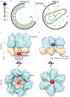
Fig. 4 ∣. Model.
a, Diagram of HIV-1 Gag, with the indicated positions of R150 (triangle; R18 in mature CA), K290 (circle; K158 in mature CA), K359 (square; K227 in mature CA). Dotted lines indicate protease cleavage sites. b, Diagram of Gag organization in immature virions (left). Following cleavage of Gag by protease (maturation), CA re-organizes to form a mature core around viral RNA (right). c-d, Surface representations of the CASP1 and CA hexamers in the immature and mature virus, respectively, with IP6 shown in its binding sites. The dramatic rearrangement of CA upon maturation is evident, as is the change of the IP6 binding site in immature and mature viruses. CANTD, blue; CACTD, orange; 6HB, purple; IP6, red.

References

    1. Gross I et al. A conformational switch controlling HIV-1 morphogenesis. EMBO J 19, 103–113 (2000). - PMC - PubMed
    1. Schur FK et al. An atomic model of HIV-1 capsid-SP1 reveals structures regulating assembly and maturation. Science 353, 506–508 (2016). - PubMed
    1. Wagner JM et al. Crystal structure of an HIV assembly and maturation switch. eLife 5, e17063 (2016). - PMC - PubMed
    1. Keller PW, Adamson CS, Heymann JB, Freed EO & Steven AC HIV-1 maturation inhibitor bevirimat stabilizes the immature Gag lattice. J. Virol 85, 1420–1428 (2011). - PMC - PubMed
    1. Wang M et al. Quenching protein dynamics interferes with HIV capsid maturation. Nat. Commun 8, 1779 (2017). - PMC - PubMed

Publication types