Review
. 2018 Sep;12(3):407-418.
doi: 10.1007/s12105-018-0908-4. Epub 2018 Aug 1.

Squamous Cell Carcinoma of the External Auditory Canal and Temporal Bone: An Update

Affiliations
Review

Squamous Cell Carcinoma of the External Auditory Canal and Temporal Bone: An Update

Benjamin M Allanson et al. Head Neck Pathol. 2018 Sep.

Abstract

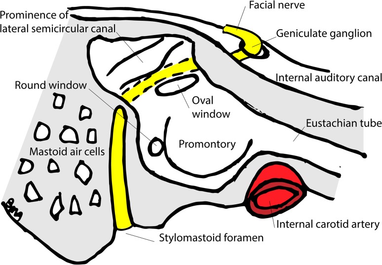
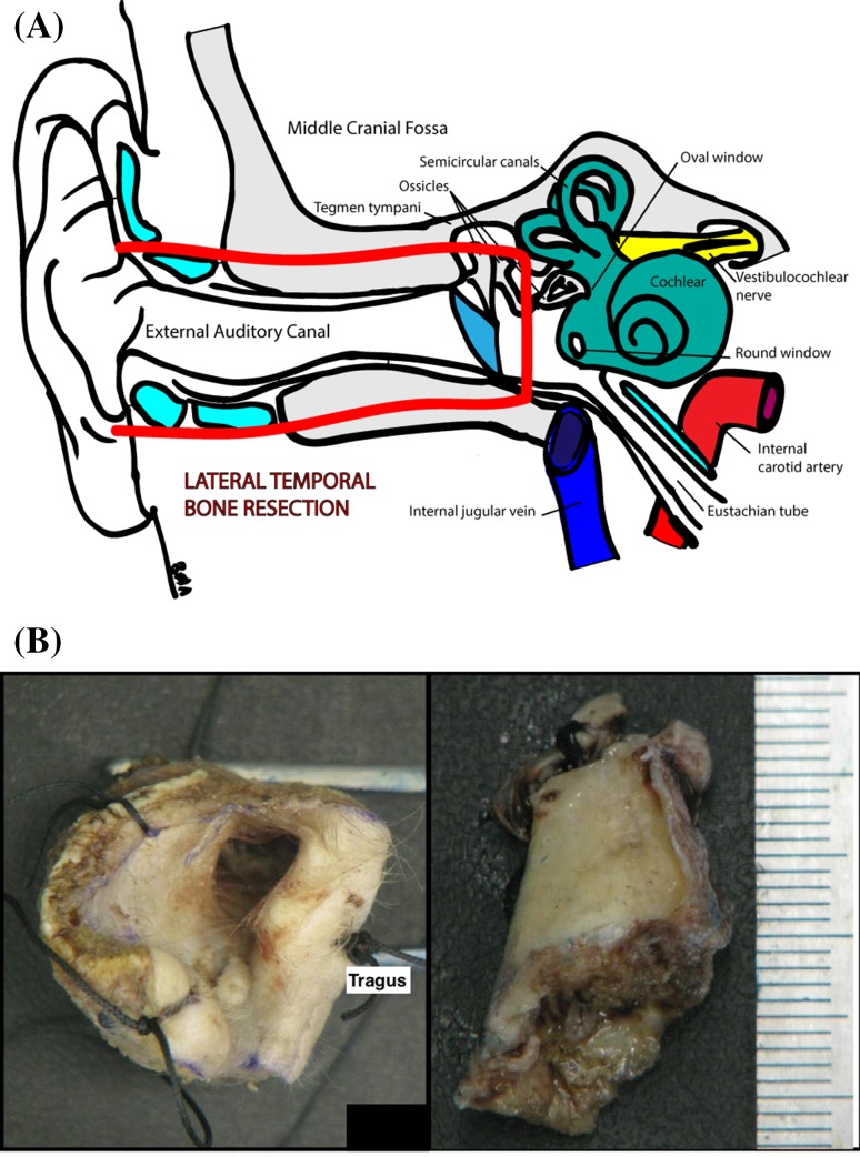
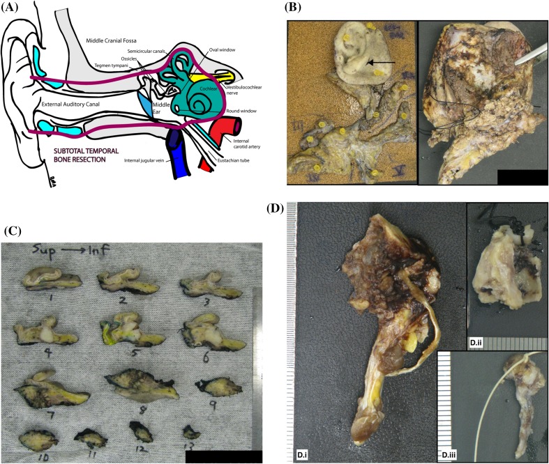
Squamous cell carcinoma (SCC) is the most common primary malignancy to affect the temporal bone, including primary cutaneous SCC of the pinna, external auditory canal, middle and inner ear. This anatomically complex region generates complicated three-dimensional specimens that can be a challenge for macroscopic and microscopic pathologic assessment. A universally accepted staging classification for these malignancies is still to be established. A brief summary of the regional anatomy, etiology and epidemiology, presentation and diagnosis, radiologic assessment and treatment follows with a review of the pathologic assessment of the different types of specimens generated and an update on staging for SCC of the temporal bone.

Keywords: External auditory canal; Macroscopic examination; Middle ear; Squamous cell carcinoma; Staging systems; Temporal bone.


Conflict of interest statement

Conflict of interest

Benjamin M. Allanson, Tsu-Hui (Hubert) Low, Jonathan R Clark, Ruta Gupta declares that they have no conflict of interest.

Research Involving Animal and Human Participants

This article does not contain any studies with human participants or animals performed by any of the authors.

Figures

Fig. 1
Fig. 1
Schematic diagram of the external auditory canal, middle and inner ear
Fig. 2
Fig. 2
Normal hematoxylin and eosin stained histology of a cartilaginous lateral one-third (inset: high power view of squamous epithelium, hair follicles and distinctive ceruminous glands) and b bony medial two-thirds of external auditory canal (inset: high power view of squamous epithelium to underlying bone)
Fig. 3
Fig. 3
Schematic diagram of the medial wall of the middle ear; representing the pathway of the facial nerve
Fig. 4
Fig. 4
Otoscopic views; a and b squamous cell carcinoma of the external auditory canal is associated with ulceration and inflammation. The edema and tissue friability makes examination painful and visibility poor
Fig. 5
Fig. 5
Hematoxylin and eosin stained histopathology of selected external ear canal malignancies; a external auditory canal with moderately differentiated squamous cell carcinoma, b poorly differentiated squamous cell carcinoma, c basal cell carcinoma (inset: high power view of nested, haphazardly arranged basaloid tumor cells with peripheral palisading), d adenoid cystic carcinoma showing a typical tubular pattern, with pseudoglandular spaces containing excess basement membrane material lined by luminal cells with dark, angular nuclei and surrounded by bland myoepithelial cells
Fig. 6
Fig. 6
Hematoxylin and eosin stained histopathology of selected benign destructive processes; a cholesterol granuloma, b cholesteotoma
Fig. 7
Fig. 7
a Schematic diagram demonstrating the anatomic structure included in a sleeve resection and b specimen photo of sleeve resection
Fig. 8
Fig. 8
a Schematic diagram demonstrating the anatomic structures included in a lateral temporal bone resection and b specimen photograph of oriented right lateral temporal bone resection with the external component (left) and excision of bony external auditory canal (right)
Fig. 9
Fig. 9
a Schematic diagram demonstrating the anatomic structures included in a subtotal temporal bone resection, b specimen photograph of contiguous pinnectomy and neck dissection oriented on a cork board (left) and posterior view of pinnectomy with underlying temporal bone (right) for metastatic squamous cell carcinoma involving a post auricular lymph node (arrow), c axial slices from superior to inferior demonstrating metastatic squamous cell carcinoma in post auricular lymph node (28 mm in maximum dimension). d (i) The styloid process, (ii) the mastoid tip, D (iii) facial nerve with suture indicating the point of exit from the stylomastoid foramen

References

    1. Stell P, McCormick M. Carcinoma of the external auditory meatus and middle ear; prognostic factors and a suggested staging system. J Laryngol Otol. 1985;99:847–850. doi: 10.1017/S0022215100097796. - DOI - PubMed
    1. The International Collaboration on Cancer Reporting. http://www.iccr-cancer.org.
    1. Snell R. Clinical anatomy by regions. 9. Philadelphia: Lippincott Williams & Wilkins; 2012.
    1. Ahn J, Park HJ, Park G, Jeong Y, Kwak H. Tympanometry and CT measurement of middle ear volumes in patients with unilateral chronic otitis media. Clin Exp Otolrhinolaryngol. 2008;1:139–142. doi: 10.3342/ceo.2008.1.3.139. - DOI - PMC - PubMed
    1. Thompson L. Update from the 4th Edition of the World Health Organization classification of head and neck tumours: tumours of the ear. Head Neck Pathol. 2017;11:78–87. doi: 10.1007/s12105-017-0790-5. - DOI - PMC - PubMed

MeSH terms

LinkOut - more resources