Review
. 2018 Sep;12(3):419-429.
doi: 10.1007/s12105-018-0915-5. Epub 2018 Aug 1.

Cholesteatoma Pearls: Practical Points and Update

Affiliations
Review

Cholesteatoma Pearls: Practical Points and Update

James T Castle. Head Neck Pathol. 2018 Sep.

Abstract

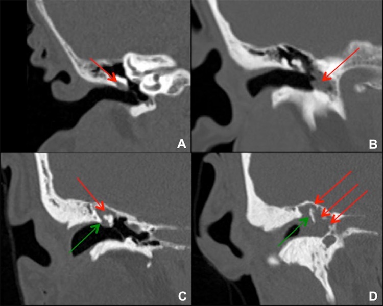
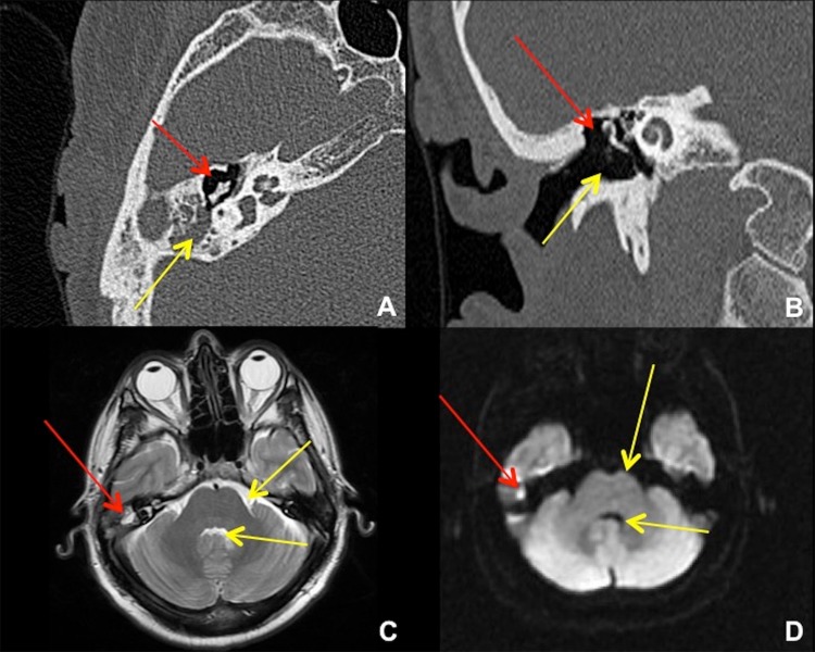
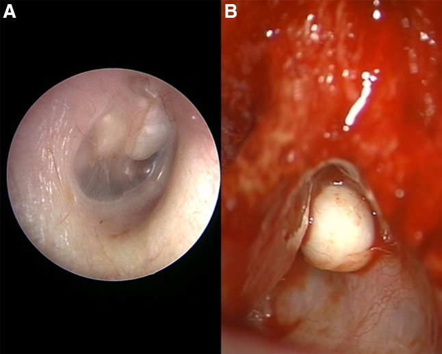
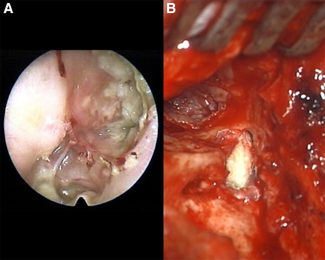
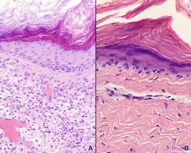
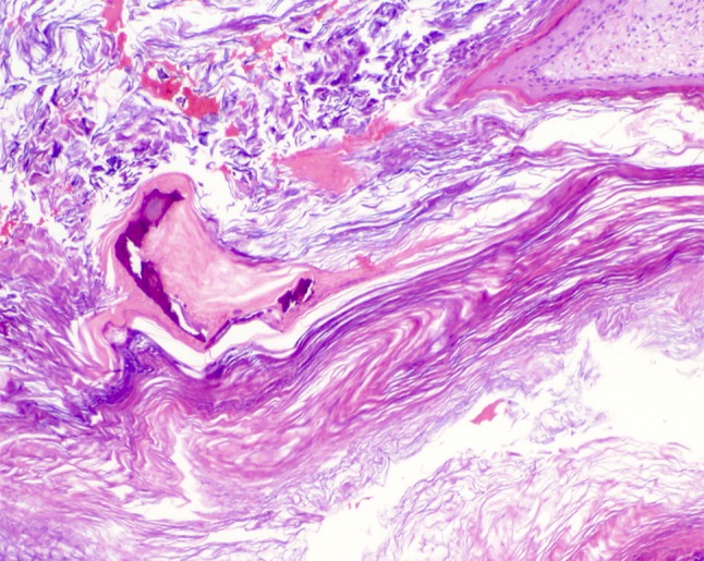
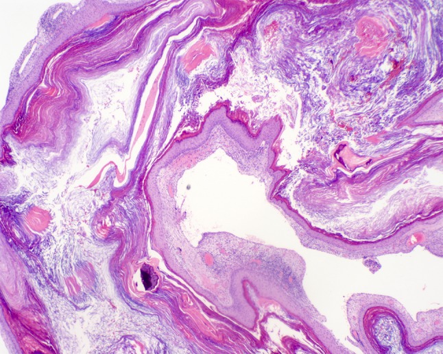
The European Academy of Otology and Neurotology in collaboration with the Japanese Otological Society (EAONO/JOS) recently produced a joint consensus document outlining the definitions, classification and staging of middle ear cholesteatoma. The goals were to provide terminologies in the description of cholesteatoma, classify cholesteatoma into distinct categories to facilitate the comparison of surgical outcomes and to provide a staging system that reflects the severity, difficulty of complete removal and restoration of normal function. Cholesteatoma is considered a benign, expanding and destructive epithelial lesion of the temporal bone that is the result of a multifactorial process. If undetected and left treated, cholesteatoma may lead to significant complications including hearing loss, temporal bone destruction and cranial invasion. Recent advances in imaging modalities have allowed for high sensitivity and specificity in identifying the presence of cholesteatoma. Despite these advances, deficiencies exist around the world with access to health care facilities meaning cholesteatoma remains a serious and challenging entity to manage whether found within the pediatric or adult population. Proper diagnosis and management of each form of cholesteatoma is achieved by a thorough understanding of the etiology, classification, clinical presentation and histology, thereby facilitating prevention, early detection and appropriate treatment.

Keywords: Acquired cholesteatoma; Cholesteatoma; Congenital cholesteatoma; Middle ear cholesteatoma.


Conflict of interest statement

Conflict of interest

James T. Castle declares that he has no conflict of interest.

Ethical Approval

This article does not contain any studies with human participants or animals performed by any of the authors.

Figures

Fig. 1
Fig. 1
Coronal images from temporal bone CTs in four different patients with right cholesteatoma. a Small cholesteatoma in Prussak’s space (red arrow) without bony erosion. This is a common site for pars flaccida retraction and acquired cholesteatoma formation. b Cholesteatoma in the left mesotympanum to hypotympanum (red arrow), which is a less common site. c Cholesteatoma in the right epitympanum (red arrow) with blunting or erosion of the right scutum (green arrow). This lesion probably started in Prussak’s space adjacent to the bony scutum. d Large cholesteatoma in the right epitympanum, mesotympanum, and hypotympanum (red arrows), with bony erosion of the scutum and malleus/ossicles (green arrow)
Fig. 2
Fig. 2
Temporal bone CT and brain MRI in 41-year-old male after transcanal endoscopic resection of a right epitympanic cholesteatoma. a Axial CT shows the resection cavity in the right Prussak’s space (red arrow), with residual cholesteatoma in the right mastoid antrum (yellow arrow). b Coronal CT shows the resection cavity at the right lateral epitympanum (red arrow) plus the surgical approach for a transcanal atticotomy (yellow arrow). An alternative approach would be via the mastoid antrum (mastoidectomy). c Axial T2-weighted MRI shows the small residual cholesteatoma (red arrow) to be of similar intensity with fluid, e.g. prepontine cistern and fourth ventricle (yellow arrows). d Axial diffusion-weighted MRI shows “restricted diffusion” in the cholesteatoma (red arrow), much brighter than free fluid (yellow arrows)
Fig. 3
Fig. 3
a Right ear with advanced congenital cholesteatoma showing involvement of the mesotympanum anterior and posterior to the malleus that appears to separate the lesion into halves. b Discrete nodule of left ear cholesteatoma at anterosuperior portion of mastoid found in the epitympanic region-aditus ad antrum, wedged between the posterior bony external auditory canal wall (at left) and the tegmen mastoideum (at right)
Fig. 4
Fig. 4
a Extensive right ear attic cholesteatoma with rupture and spilled keratinaceous contents. b Surgical entry via mastoidectomy approach where a tympanic membrane retraction pocket formed a sac into the mastoid cavity and eroded lateral canal wall. The top left of photo looks down the ear canal to the eardrum
Fig. 5
Fig. 5
a Abundant granulation tissue peripheral to the squamous epithelial lining comprises the perimatrix. Note that rete peg formation is absent but a prominent granular cell layer is present. b Dense fibrous connective tissue adjacent to the epithelium is present with a chronic inflammatory cell infiltrate found in place of granulation tissue
Fig. 6
Fig. 6
Irregular acellular basophilic calcifications that may represent eroded ossicles or bone of the middle ear canal are seen in conjunction with desquamated keratin of the cystic lumen
Fig. 7
Fig. 7
Cystic formation of bland squamous epithelium surrounding abundant layers of laminated, desquamated and anucleate keratin filling the cystic luminal area

References

    1. Yung M, Tono T, Olszewska E, et al. EAONO/JOS Joint consensus statements on the definitions, classification and staging of middle ear cholesteatoma. J Int Adv Otol. 2017;13:1–8. doi: 10.5152/iao.2017.3363. - DOI - PubMed
    1. Müller J. Ueber den feineren bau und die formen der krankhaften geschwulste. Berlin, G Reimer. 1838. Folio.
    1. De Verney JG. Traité de l’organie de l’ouïe. Paris: E. Michallet; 1683.
    1. Aquino JE, Cruz Filho NA, de Aquino JN. Epidemiology of middle ear and mastoid cholesteatomas: study of 1146 cases. Braz J Otorhinolaryngol. 2011;77:341–347. doi: 10.1590/S1808-86942011000300012. - DOI - PMC - PubMed
    1. Bennett M, Warren F, Jackson GC. Congenital cholesteatoma: theories, facts and 53 patients. Otolaryngol Clin N Am. 2006;39:1081–1094. doi: 10.1016/j.otc.2006.08.001. - DOI - PubMed

LinkOut - more resources