

Selected Giant Cell Rich Lesions of the Temporal Bone
- PMID: 30069841
- PMCID: PMC6081287
- DOI: 10.1007/s12105-018-0906-6
Selected Giant Cell Rich Lesions of the Temporal Bone
Abstract
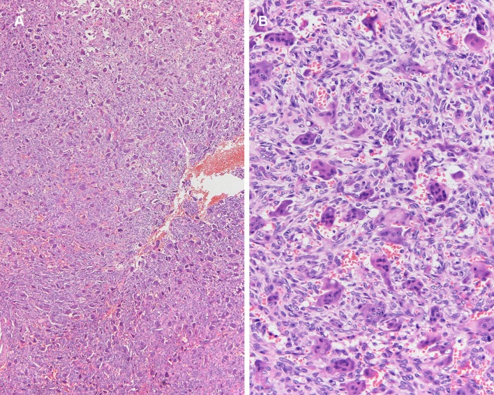
Giant cell rich lesions of the temporal bone encompass a wide spectrum of disease that includes infectious, reactive, and neoplastic processes. When dealing with any lesion that can potentially involve bone, it is important to understand both the clinical presentation and to correlate the histologic findings with the radiologic imaging. This review discusses the clinical, the pathologic features including the differential diagnosis, and the treatment of some of the more commonly encountered giant cell rich entities in this region.
Keywords: Chondroblastoma; Giant cell reparative granuloma; Langerhans cell histiocytosis; Sarcoidosis; Temporal bone; Tenosynovial giant cell tumor.
Figures

References
-
- Yu RC, Chu C, Buluwela L, Chu AC. Clonal proliferation of Langerhans cells in Langerhans cell histiocytosis. Lancet. 1994;343(8900):767–768. - PubMed
-
- Willman CL, Busque L, Griffith BB, Favara BE, McClain KL, Duncan MH, et al. Langerhans’-cell histiocytosis (histiocytosis X)--a clonal proliferative disease. N Engl J Med. 1994;331(3):154–160. - PubMed
-
- Coppes-Zantinga A, Egeler RM. The Langerhans cell histiocytosis X files revealed. Br J Haematol. 2002;116(1):3–9. - PubMed
-
- Hicks J, Flaitz CM. Langerhans cell histiocytosis: current insights in a molecular age with emphasis on clinical oral and maxillofacial pathology practice. Oral Surg Oral Med Oral Pathol Oral Radiol Endod. 2005;100(2 Suppl):S42–S66. - PubMed
-
- Abla O, Egeler RM, Weitzman S. Langerhans cell histiocytosis: current concepts and treatments. Cancer Treat Rev. 2010;36(4):354–359. - PubMed
Publication types
MeSH terms
LinkOut - more resources
Full Text Sources
Other Literature Sources
