

Dendrimer-N-acetyl-L-cysteine modulates monophagocytic response in adrenoleukodystrophy
- PMID: 30069915
- PMCID: PMC6454885
- DOI: 10.1002/ana.25303
Dendrimer-N-acetyl-L-cysteine modulates monophagocytic response in adrenoleukodystrophy
Abstract
Objective: X-linked adrenoleukodystrophy (ALD) is a neurodegenerative disorder due to mutations in the peroxisomal very long-chain fatty acyl-CoA transporter, ABCD1, with limited therapeutic options. ALD may manifest in a slowly progressive adrenomyeloneuropathy (AMN) phenotype, or switch to rapid inflammatory demyelinating cerebral disease (cALD), in which microglia have been shown to play a pathophysiological role. The aim of this study was to determine the role of patient phenotype in the immune response of ex vivo monophagocytic cells to stimulation, and to evaluate the efficacy of polyamidoamine dendrimer conjugated to the antioxidant precursor N-acetyl-cysteine (NAC) in modulating this immune response.
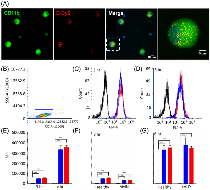
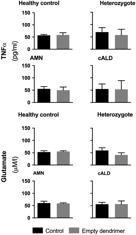
Methods: Human monophagocytic cells were derived from fresh whole blood, from healthy (n = 4), heterozygote carrier (n = 4), AMN (n = 7), and cALD (n = 4) patients. Cells were exposed to very long-chain fatty acids (VLCFAs; C24:0 and C26:0) and treated with dendrimer-NAC (D-NAC).
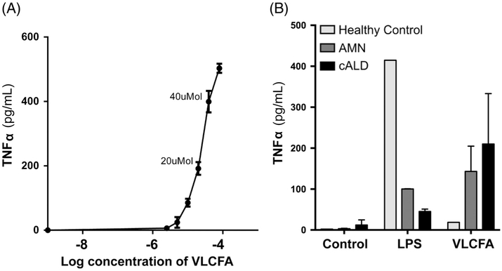
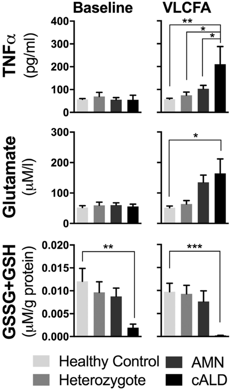
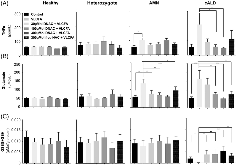
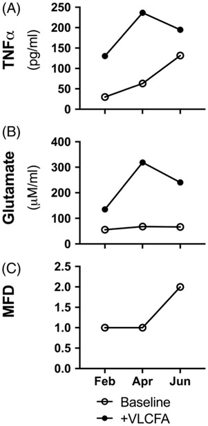
Results: Ex vivo exposure to VLCFAs significantly increased tumor necrosis factor α (TNFα) and glutamate secretion from cALD patient macrophages. Additionally, a significant reduction in total intracellular glutathione was observed in cALD patient cells. D-NAC treatment dose-dependently reduced TNFα and glutamate secretion and replenished total intracellular glutathione levels in cALD patient macrophages, more efficiently than NAC. Similarly, D-NAC treatment decreased glutamate secretion in AMN patient cells.
Interpretation: ALD phenotypes display unique inflammatory profiles in response to VLCFA stimulation, and therefore ex vivo monophagocytic cells may provide a novel test bed for therapeutic agents. Based on our findings, D-NAC may be a viable therapeutic strategy for the treatment of cALD. Ann Neurol 2018;84:452-462.
© 2018 American Neurological Association.
Conflict of interest statement
Potential Conflicts of Interest
B.Tu., S.Kan., R.K., and A.F. report a patent application US20170119899 issued October 16, 2018 for the use of dendrimer technologies described in this paper. The patent will be owned by those authors. At the time of publication, the patent is licensed to Ashvattha. S.Kan. and R.K. are the cofounders of the companies Ashvattha and Orpheris, which focus on therapies with the dendrimer platform. S.Kan. and R.K. are cofounders and members of the board of directors and own shares in Ashvattha and Orpheris, companies that are translating and commercializing the dendrimer platform.
Figures
References
-
- Moser HW, Mahmood A, Raymond GV. X-linked adrenoleukodystrophy. Nat Clin Pract Neurol 2007;3:140–151. - PubMed
-
- Kemp S, Huffnagel IC, Linthorst GE, et al. Adrenoleukodystrophy—neuroendocrine pathogenesis and redefinition of natural history. Nat Rev Endocrinol 2016;12:606–615. - PubMed
-
- Eichler FS, Ren J-Q, Cossoy M, et al. Is microglial apoptosis an early pathogenic change in cerebral X-linked adrenoleukodystrophy? Ann Neurol 2008;63:729–742. - PubMed
Publication types
MeSH terms
Substances
Grants and funding
LinkOut - more resources
Full Text Sources
Other Literature Sources
