. 2018 Oct;110(1):128-142.
doi: 10.1111/mmi.14093. Epub 2018 Oct 2.

In silico discovery of small molecules that inhibit RfaH recruitment to RNA polymerase

Affiliations

In silico discovery of small molecules that inhibit RfaH recruitment to RNA polymerase

Dmitri Svetlov et al. Mol Microbiol. 2018 Oct.

Abstract

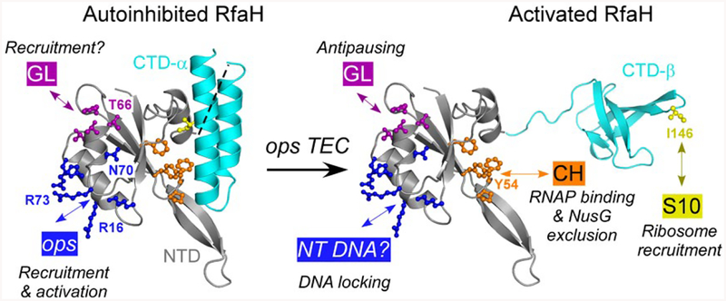
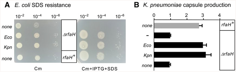
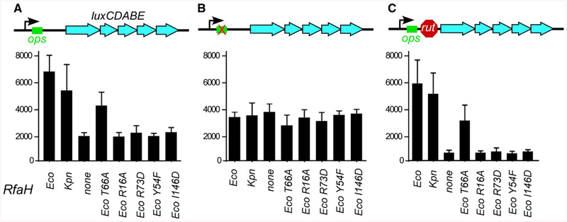
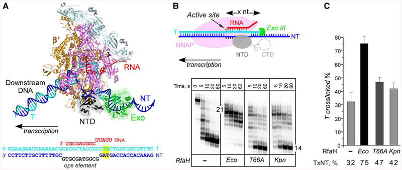
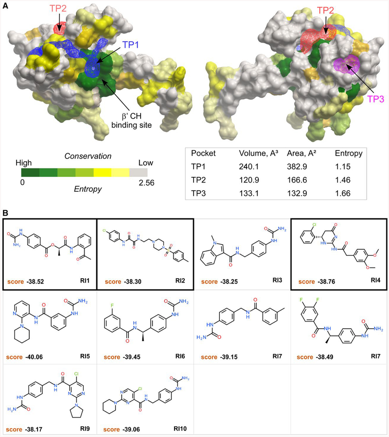
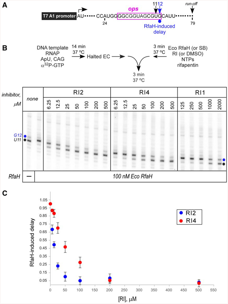
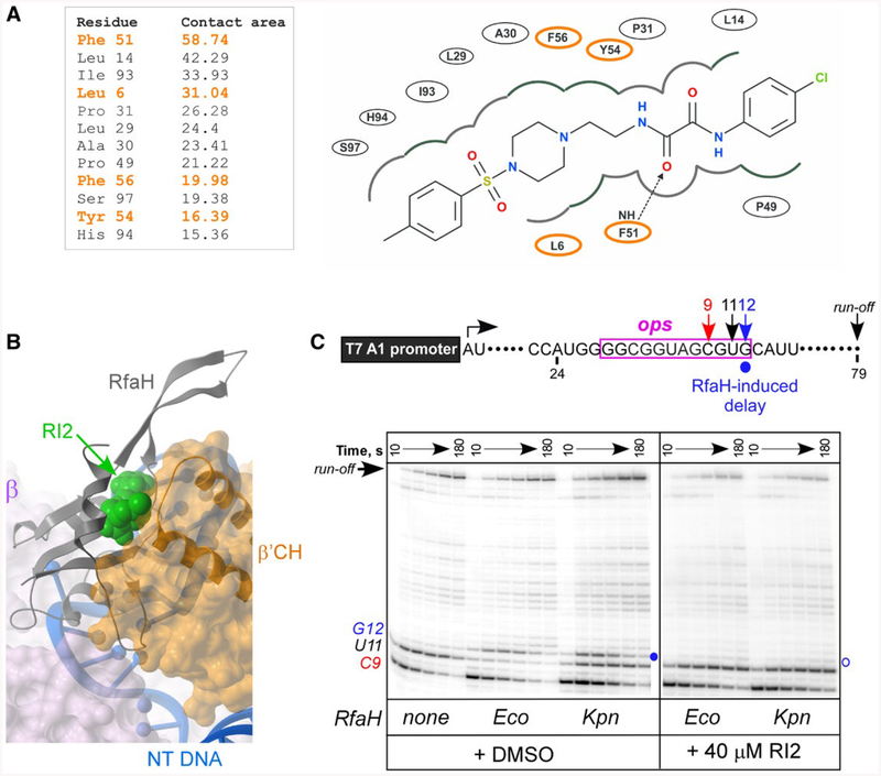
RfaH is required for virulence in several Gram-negative pathogens including Escherichia coli and Klebsiella pneumoniae. Through direct interactions with RNA polymerase (RNAP) and ribosome, RfaH activates the expression of capsule, cell wall and pilus biosynthesis operons by reducing transcription termination and activating translation. While E. coli RfaH has been extensively studied using structural and biochemical approaches, limited data are available for other RfaH homologs. Here we set out to identify small molecule inhibitors of E. coli and K. pneumoniae RfaHs. Results of biochemical and functional assays show that these proteins act similarly, with a notable difference between their interactions with the RNAP β subunit gate loop. We focused on high-affinity RfaH interactions with the RNAP β' subunit clamp helices as a shared target for inhibition. Among the top 10 leads identified by in silico docking using ZINC database, 3 ligands were able to inhibit E. coli RfaH recruitment in vitro. The most potent lead was active against both E. coli and K. pneumoniae RfaHs in vitro. Our results demonstrate the feasibility of identifying RfaH inhibitors using in silico docking and pave the way for rational design of antivirulence therapeutics against antibiotic-resistant pathogens.


Conflict of interest statement

Conflict of interest

The Authors declare no conflicts of interest.

Figures

Fig. 1.
Fig. 1.
Key residues in Eco RfaH. In the autoinhibited state, the NTD (gray) and CTD (α-helices; cyan) interact to bury the NTD residues that bind to the β’CH domain (orange). In the active state, the domains are connected by a (modeled) flexible linker; the NTD and the β-barrel CTD bind to RNAP and S10 respectively. In both states, the NTD can interact with the nontemplate (NT) ops DNA element (blue) and the βGL (dark magenta).
Fig. 2.
Fig. 2.
Plasmid-encoded Eco and Kpn RfaH complement rfaH deletions in E. coli and K. pneumoniae. A. Dilutions of exponentially growing cultures of MG1655ΔrfaH strain transformed with plasmids expressing Eco RfaH, Kpn RfaH, or a control vector were plated on LB-chloramphenicol (left) or LB-Cm supplemented with 0.5% SDS and 0.2 mM IPTG (right) and incubated at 37°C overnight. A representative set from three independent experiments is shown B. Relative capsule production in K. pneumoniae TOP52 or TOP52ΔrfaH strains transformed with plasmids containing Eco RfaH, Kpn RfaH, or no insert. Data are combined from three independent experiments, normalized to TOP52ΔrfaH without an RfaH plasmid, and error bars represent standard deviation.
Fig. 3.
Fig. 3.
Reporter assays in K. pneumoniae. Plasmids encoding wild-type RfaH proteins or Eco RfaH variants with single-residue substitutions under the control of Ptrc promoter were co-transformed into TOP52ΔrfaH strain with reporter vectors containing the Photorhabdus luminescens luxCDABE operon under the control of PBAD promoter, with ops and rut elements in the leader region as indicated in the schematics. The results are expressed as luminescence corrected for the cell densities of individual cultures. Data are combined from three independent experiments and error bars represent standard deviation.
Fig. 4.
Fig. 4.
Probing RfaH-DNA interactions. A. A model of the RfaH-bound TEC. RNAP α (pale cyan), β (magenta) and β’ (orange) subunits, RfaH-NTD (gray), nucleic acids and Exo (green) are shown as cartoons. To provide an unobstructed view of the RfaH-NTD and the exposed nontemplate DNA, the EC is shown inan orientation that is opposite to the conventional left-to-right direction of transcription. The ops TEC scaffold used in these experiments is shown below, with nucleic acid chains colored and oriented as in the model; the ops element is in black. The upstream TA cross-linking motif is highlighted in yellow. B. Footprinting of the upstream RNAP boundary. The template strand DNA was 5’-end labeled with [γ32P]-ATP. After the addition of Exo III, aliquots were quenched at the indicated times (0 represents an untreated DNA control) and analyzed on a 12% denaturing gel; a representative of three independent experiments is shown. Numbers indicate the distance from the RNAP active site (yellow circle). C. Probing the upstream fork junction by cross-linking with 8-MP. TECs were supplemented with 100 nM Eco or 250 nM Kpn RfaH (where indicated) and illuminated with the 365 nm UV light. Fractions of the cross-linked DNA were determined after analysis on denaturing gels. Error bars indicate the SDs of triplicate measurements. See also Fig. S2.
Fig. 5.
Fig. 5.
Tentative pockets on the RfaH-NTD and structures of potential inhibitors. A. Three tentative pockets (TP1, blue; TP2, red; and TP3, magenta) identified by ICMPocketFinder tool in ICM-Pro v3.8–6a are shown as transparent meshes; the volume and area data are shown in the table below. The RfaH-NTD is shown as a molecular surface where residues are colored by the alignment conservation Entropies (see Methods and Materials), with highly conserved (low Entropy) residues shown in green. The Entropy of each pocket was calculated as the average Entropy of residues around the pocket. B. Structures and docking scores of the top 10 hits from virtual ligand screening predicted to bind to TP1. Three molecules that show inhibitory activity against RfaH are indicated by thick borders.
Fig. 6.
Fig. 6.
Inhibition of Eco RfaH recruitment by RIs. A. Transcript generated from the T7A1 promoter on a linear DNA template; transcription start site (a bent arrow), ops element (magenta box), pause sites and transcript end are indicated on top. B. Halted A24 TECs were formed as described in Materials and Methods. Elongation was restarted upon addition of NTPs and rifapentin in the presence of Eco RfaH (100 nM) preincubated with increasing concentrations of RI 1, 2 or 4. Aliquots were withdrawn at selected times and analyzed on a 10% denaturing gel. Positions of the paused and run-off transcripts are indicated; the position of the RfaH-induced RNAP pause at G12 is indicated with a circle. C. The fraction of G12 RNA was quantified as a function of RI concentration and corrected for levels observed in the absence of RfaH; the G12 RNA in the absence of RI (DMSO control) was defined as 1. The results of triplicate measurements for RI2 and RI4 are shown; errors are ± SD. Assays with RI1 were also performed in triplicates, but the observed inhibition was too weak to accurately determine the apparent IC50.
Fig. 7.
Fig. 7.
RI2 is predicted to block RfaH interactions with the β’CH domain. A. RfaH residues interacting with RI2 in the predicted binding pose. Residues indicated in orange interact with β’CH of RNAP (Kang et al., 2018). Left, contact areas (Å2) of the RfaH-NTD residues interacting with RI2. Right, a 2D interaction diagram of RfaH-NTD and RI2 in the predicted model. The dashed line with an arrow represents a hydrogen bond between residues and RI2. B. Superposition of the modeled RfaH-NTD/RI2 complex and the cryo-electron microscopy structure (PDB ID: 6C6T) of the RfaH/TEC complex (Kang et al., 2018) using the RfaH-NTD backbone. Binding of RI2 (green) is incompatible with the β’CH (orange). C. Effects of RI2 on RfaH recruitment at the ops site. Halted A24 TECs were formed as described in Materials and Methods. Elongation was restarted upon addition of NTPs and rifapentin in the presence of Eco or Kpn RfaH (100 nM) and RI2 (or DMSO). Aliquots were withdrawn at selected times and analyzed on a 10% denaturing gel. Positions of the paused and run-off transcripts are indicated; the position of the RfaH-induced RNAP pause at G12 is indicated with a circle.

References

    1. Abagyan R and Kufareva I (2009) The flexible pocketome engine for structural chemogenomics. Methods in Molecular Biology, 575, 249–279. - PMC - PubMed
    1. Abagyan R, Raush E, Totrov M and Orry A (2017) ICM Manual v3.8–6. San Diego, CA: Molsoft LCC.
    1. Abagyan R, Totrov M and Kuznetsov D (1994) ICM – a new method for protein modeling and design: applications to docking and structure prediction from the distorted native conformation. Journal of Computational Chemistry, 15, 488–506.
    1. An J, Totrov M and Abagyan R (2005) Pocketome via comprehensive identification and classification of ligand binding envelopes. Molecular & Cellular Proteomics, 4, 752–761. - PubMed
    1. Artsimovitch I and Landick R (2002) The transcriptional regulator RfaH stimulates RNA chain synthesis after recruitment to elongation complexes by the exposed non-template DNA strand. Cell, 109, 193–203. - PubMed

Publication types

MeSH terms