

Removal of large impacted foreign body from the base of the skull through submandibular access: A multidisciplinary approach
- PMID: 30071376
- PMCID: PMC6080633
- DOI: 10.1016/j.ijscr.2018.07.012
Removal of large impacted foreign body from the base of the skull through submandibular access: A multidisciplinary approach
Abstract
Introduction: This report describes the removal of a missed impacted large piece of a glass that reaches the infra-temporal region after a traumatic injury at the submandibular area.
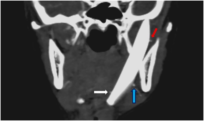
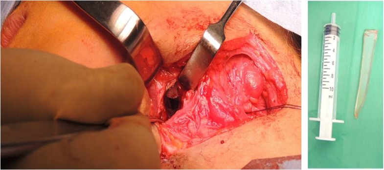
Case presentation: A nine year-old patient presented with a limited mouth opening (0.5 cm). Initial examination showed a scar of an old trauma in the submandibular area two months prior to presentation. The radiographic study showed a large knife-shaped foreign body with its tip at the infra-temporal region, and its base at the submandibular region. Further multi-slice computed tomography with angiography was done that showed close proximity of the foreign body to the branches of the external carotid artery; maxillary, lingual and facial branches. The foreign body was removed via extra-oral approach through the old scar of the past injury under general anesthesia. Dissection, exposure of the external carotid artery and preparing it for emergency ligation, were done before extraction of the foreign body. The patient's mouth opening increased to 2.5 cm without any complications.
Conclusion: Cut wounds in the face should not be repaired in the primary care without detailed history, systematic examination and proper investigations.
Keywords: External carotid artery; Foreign body; Infra-temporal cone beam computed tomography; Skull base; Submandibular.
Copyright © 2018. Published by Elsevier Ltd.
Figures


References
-
- Ueeck B.A. Penetrating injuries to the face: delayed versus primary treatment—considerations for delayed treatment. J. Oral Maxillofac. Surg. 2007;65(6):1209–1214. - PubMed
-
- Eggers G., Haag C., Hassfeld S. Image-guided removal of foreign bodies. Br. J. Oral Maxillofac. Surg. 2005;43(5):404–409. - PubMed
-
- Mohanavalli S., David J.J., Gnanam A. Rare foreign bodies in oro-facial regions. Indian J. Dent. Res. 2011;22(5):713–715. - PubMed
-
- Santos Tde S. Impacted foreign bodies in the maxillofacial region-diagnosis and treatment. J. Craniofac. Surg. 2011;22(4):1404–1408. - PubMed
-
- Agha R.A., Fowler A.J., Saetta A., Barai I., Rajmohan S., Orgill D.P., for the SCARE Group The SCARE statement: consensus-based surgical case report guidelines. Int. J. Surg. 2016;27:187–189. - PubMed
LinkOut - more resources
Full Text Sources
Other Literature Sources
Miscellaneous
