

Metastatic Complications of Cancer Involving the Central and Peripheral Nervous Systems
- PMID: 30072072
- PMCID: PMC6082424
- DOI: 10.1016/j.ncl.2018.04.011
Metastatic Complications of Cancer Involving the Central and Peripheral Nervous Systems
Abstract
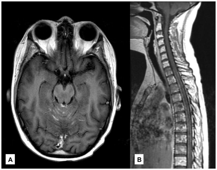
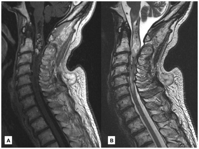
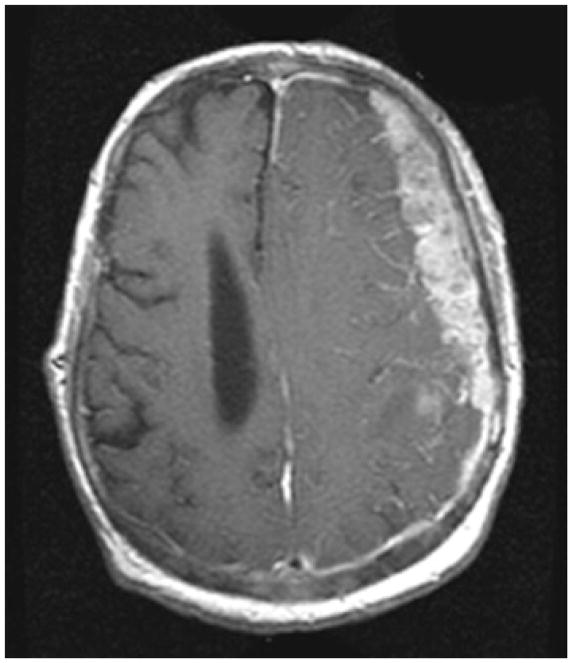
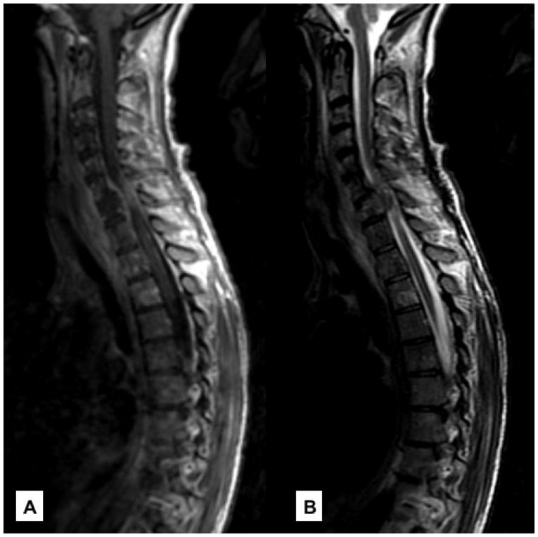
Neurologic complications of cancer may involve both the central nervous system and peripheral nervous system manifesting as brain, leptomeningeal, intramedullary, intradural, epidural, plexus, and skull base metastases. Excluding brain involvement, neurologic complications affecting these other sites are relatively infrequent, but collectively they affect more than 25% of patients with metastatic cancer causing significant morbidity and mortality. Early diagnosis and intervention optimize quality of life and improve survival.
Keywords: CNS; Cancer; Epidural cord compression; Leptomeningeal; Neurologic complications; PNS; Skull base and nerve plexuses.
Copyright © 2018 Elsevier Inc. All rights reserved.
Conflict of interest statement
Dr. Joe Mendez has no conflict of interest
Dr. Lisa DeAngelis has no conflict of interest
Figures
References
-
- DeAngelis LM, Posner JB, Posner JB. Neurologic complications of cancer. 2. Oxford ; New York: Oxford University Press; 2009.
-
- Norris LK, Grossman SA, Olivi A. Neoplastic meningitis following surgical resection of isolated cerebellar metastasis: A potentially preventable complication. J Neuro-Oncol. 1997;32(3):215–223. - PubMed
-
- Glantz MJ, Cole BF, Glantz LK, et al. Cerebrospinal fluid cytology in patients with cancer: minimizing false-negative results. Cancer. 1998;82(4):733–739. - PubMed
-
- Rogers LR, Duchesneau PM, Nunez C, et al. Comparison of Cisternal and Lumbar Csf Examination in Leptomeningeal Metastasis. Neurology. 1992;42(6):1239–1241. - PubMed
-
- Bromberg JE, Breems DA, Kraan J, et al. CSF flow cytometry greatly improves diagnostic accuracy in CNS hematologic malignancies. Neurology. 2007;68(20):1674–1679. - PubMed
Publication types
MeSH terms
Grants and funding
LinkOut - more resources
Full Text Sources
Other Literature Sources
Medical
