. 2018 Sep 21;293(38):14823-14838.
doi: 10.1074/jbc.RA118.004773. Epub 2018 Aug 2.

Roles of distal aspartate and arginine of B-class dye-decolorizing peroxidase in heterolytic hydrogen peroxide cleavage

Affiliations

Roles of distal aspartate and arginine of B-class dye-decolorizing peroxidase in heterolytic hydrogen peroxide cleavage

Vera Pfanzagl et al. J Biol Chem. .

Abstract

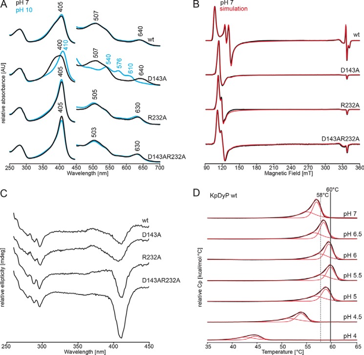
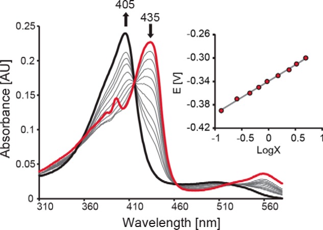
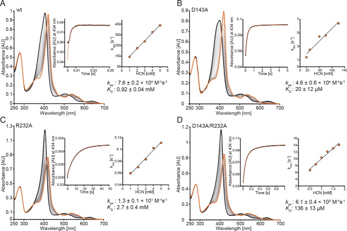
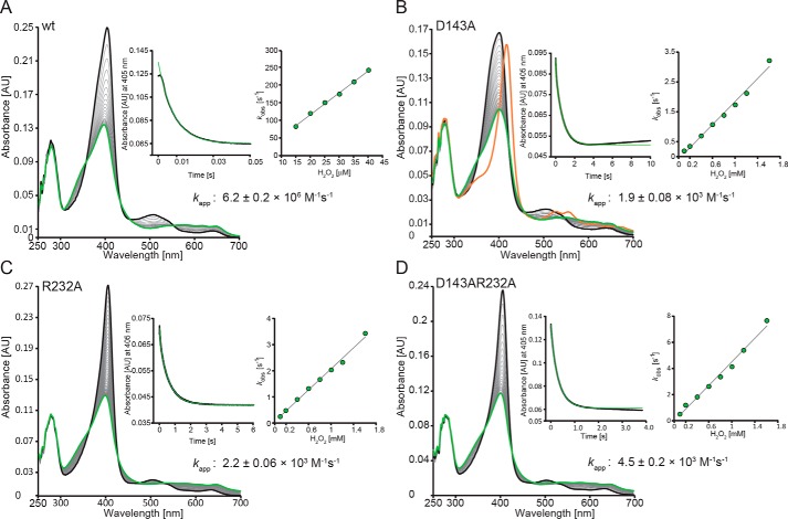
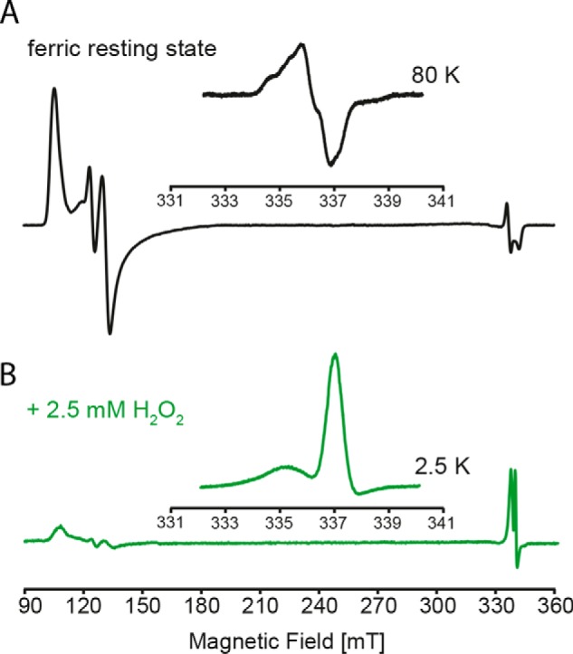
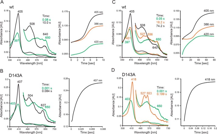
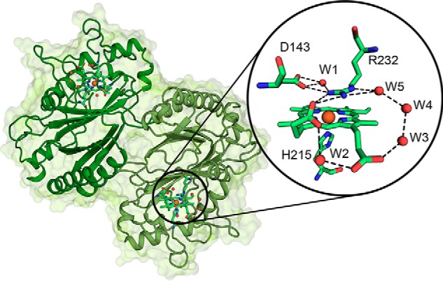
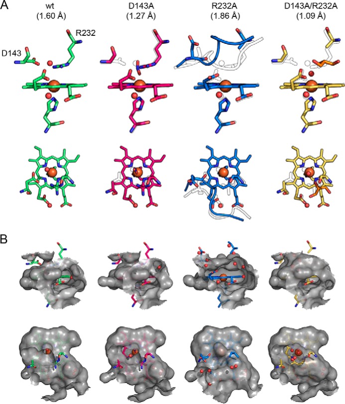
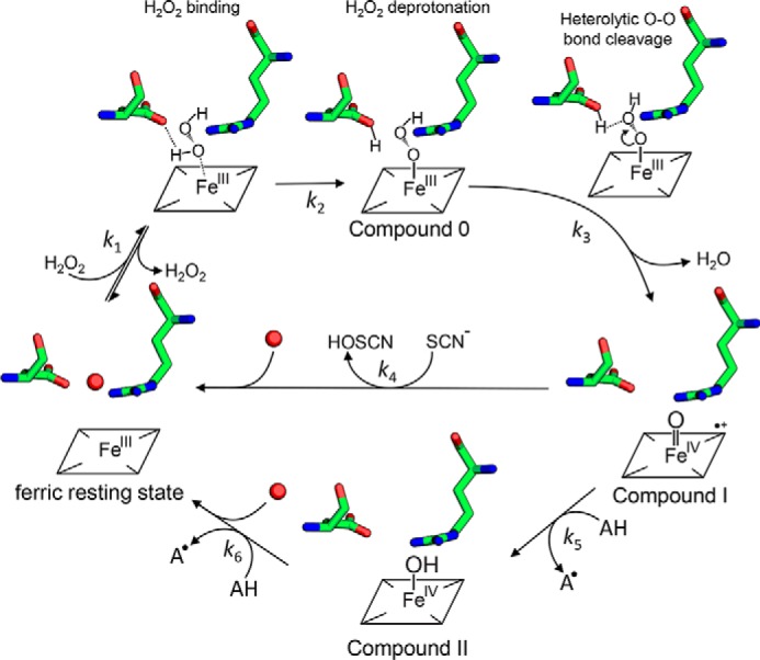
Dye-decolorizing peroxidases (DyPs) represent the most recently classified hydrogen peroxide-dependent heme peroxidase family. Although widely distributed with more than 5000 annotated genes and hailed for their biotechnological potential, detailed biochemical characterization of their reaction mechanism remains limited. Here, we present the high-resolution crystal structures of WT B-class DyP from the pathogenic bacterium Klebsiella pneumoniae (KpDyP) (1.6 Å) and the variants D143A (1.3 Å), R232A (1.9 Å), and D143A/R232A (1.1 Å). We demonstrate the impact of elimination of the DyP-typical, distal residues Asp-143 and Arg-232 on (i) the spectral and redox properties, (ii) the kinetics of heterolytic cleavage of hydrogen peroxide, (iii) the formation of the low-spin cyanide complex, and (iv) the stability and reactivity of an oxoiron(IV)porphyrin π-cation radical (Compound I). Structural and functional studies reveal that the distal aspartate is responsible for deprotonation of H2O2 and for the poor oxidation capacity of Compound I. Elimination of the distal arginine promotes a collapse of the distal heme cavity, including blocking of one access channel and a conformational change of the catalytic aspartate. We also provide evidence of formation of an oxoiron(IV)-type Compound II in KpDyP with absorbance maxima at 418, 527, and 553 nm. In summary, a reaction mechanism of the peroxidase cycle of B-class DyPs is proposed. Our observations challenge the idea that peroxidase activity toward conventional aromatic substrates is related to the physiological roles of B-class DyPs.

Keywords: Compound I; Compound II; Klebsiella pneumonia; X-ray crystallography; electron paramagnetic resonance (EPR); enzyme kinetics; heme; heme peroxidase; oxoiron; pre-steady-state kinetics; site-directed mutagenesis.


Conflict of interest statement

The authors declare that they have no conflicts of interest with the contents of this article

Figures

Figure 1.
Figure 1.
Spectral properties and thermal stabilities of WT KpDyP and the variants D143A, R232A, and D143A/R232A. A, UV-visible spectra of WT KpDyP, D143A, R232A, and D132A/R232A in 50 mm phosphate buffer, pH 7.0 (black), and in 50 mm borate buffer pH 10.0 (blue). B, low temperature X-band CW EPR spectra of WT KpDyP and variants in 50 mm phosphate buffer, pH 7.0 (black), and their corresponding spectral simulations (red). C, CD spectra of WT KpDyP and variants in 50 mm phosphate buffer, pH 7.0, in the near-UV region (260–450 nm). D, DSC thermograms of WT KpDyP between pH 4.0 and 7.0 (black). The corresponding fits using a non-two-state transition model are depicted in red. The highest Tm values (pH 5.5) are highlighted with gray lines.
Figure 2.
Figure 2.
Spectroelectrochemical titration of WT KpDyP. Shown are electronic absorption spectra of WT KpDyP at different applied potentials. The bold black spectrum represents fully oxidized, ferric enzyme (Soret maximum at 405 nm), and the red spectrum represents fully reduced (Soret maximum at 435 nm) protein. The inset shows the corresponding Nernst plot, where x represents (AλredMaxAλred)/(AλoxMaxAλox).
Figure 3.
Figure 3.
Kinetics of cyanide binding to ferric WT KpDyP and the variants D143A, R232A, and D143A/R232A at pH 7.0. Spectral conversion of high-spin ferric protein (black spectrum) to the corresponding low-spin cyanide complex (orange spectrum) is shown. A, WT; B, D143A; C, R232A; D, D143AR232A. The insets show representative time traces (black) and fits (orange) at 434 nm (WT KpDyP, 5 mm; D143A, 100 μm; R232A, 5 mm; D143A/R232A, 1.5 mm). Additionally, insets show the corresponding linear plots of kobs versus cyanide concentration.
Figure 4.
Figure 4.
Kinetics of Compound I formation of WT KpDyP and the variants D143A, R232A, and D143A/R232A at pH 7.0. Spectral transition upon the reaction of 2 μm WT (A), D143A (B), R232A (C), and D143A/R232A (D) with 20 μm (WT) or 800 μm (variants) hydrogen peroxide is shown. Insets depict typical time traces at 405 nm (black) and the corresponding single- or double-exponential fits (green). Additionally, insets show the corresponding linear plots of kobs versus hydrogen peroxide concentration.
Figure 5.
Figure 5.
Low temperature CW EPR of WT KpDyP and Compound I. A, X-band CW EPR spectrum of WT KpDyP. The inset shows the organic radical signature of the resting state at 80 K. B, X-band CW EPR spectrum of KpDyP Compound I formed by the addition of a 5-fold stoichiometric excess of hydrogen peroxide. The inset shows the porphyryl radical signature observed at 2.5 K.
Figure 6.
Figure 6.
Reaction of WT KpDyP and D143A Compound I with thiocyanate and serotonin. A and C, reaction of WT Compound I with thiocyanate (A) and serotonin (C) at pH 7.0, followed by the sequential stopped-flow mode. Compound I (green spectrum) was preformed by mixing 2 μm ferric KpDyP with 4 μm H2O2 in 50 mm phosphate buffer, pH 7.0. After a 100-ms delay time, 1 mm thiocyanate (A) or 50 μm serotonin (C) was added. B and D, reaction of D143A Compound I with thiocyanate (B) and serotonin (D) at pH 7.0, followed by the sequential stopped-flow mode. Compound I (green spectrum) was preformed by mixing 2 μm ferric D143A with 1 mm μm H2O2 in 50 mm phosphate buffer, pH 7.0. After a 3000-ms delay time, 10 μm thiocyanate (A) or 1 mm serotonin (C) was added. Ferric spectra are shown in black, and oxoiron(IV)-type Compound II spectra are shown in orange. Relevant absorption maxima and time points of selection of the spectra are depicted.
Figure 7.
Figure 7.
Overall and active-site crystal structure of WT KpDyP. The dimeric structure of KpDyP is shown as a cartoon (green). Heme prosthetic groups are depicted as sticks, and the heme iron is shown as an orange sphere. The round inset shows a detailed view of the active-site amino acid residues Asp-143 and Arg-232 (distal) and His-215 (proximal) and the heme b moiety as a stick representation. Hydrogen-bonding networks involving the distal residues and the heme b propionates are shown as dashed lines, and water molecules (W) and heme iron are depicted as red and orange spheres, respectively.
Figure 8.
Figure 8.
Comparison of the active-site architecture of WT KpDyP and the variants D143A, R232A, and D143A/R232A. A, representation of the active-site residues Asp-143, Arg-232, and His-215 and the heme b moiety shown from the front (first row) and top (second row). Relevant water molecules are shown as red spheres, nitrite (D143A structure) is shown in blue, and glycerol (D132A/R232A structure) is shown in orange. In all mutant structures, the WT structure is depicted as a black outline for comparison. Additionally, the backbones of residues 141–148 are shown in the R232A structure together with the respective WT conformation (black outline). B, surface of the active site and access channels shown in a 6-Å radius from the heme iron colored in gray (semitransparent), shown from the front (first row) to visualize the surface-accessible propionates and from the top (second row) to show the distal access channel.
Figure 9.
Figure 9.
Proposed reaction mechanism for Compound 0, Compound I, and Compound II formation in B-class DyPs. Hydrogen peroxide enters the narrow distal heme access channel, binds (k1), and is deprotonated by Asp-143 (Compound 0 formation). Heterolytic cleavage of the O–O bond (k3) forms Compound I (oxoiron(IV) porphyrin radical) (k3), which is reduced by either two-electron donors like thiocyanate (SCN) directly to the ferric resting state, thereby producing hypothiocyanite (HOSCN) (k4), or by one-electron donors like serotonin (AH) to Compound II (oxoiron(IV) species) (k5), thereby producing the corresponding radical (A). Finally, a second one-electron donor (AH) reduces Compound II to the ferric resting state (k6).

References

    1. Kim S. J., and Shoda M. (1999) Decolorization of molasses and a dye by a newly isolated strain of the fungus Geotrichum candidum Dec 1. Biotechnol. Bioeng. 62, 114–119 10.1002/(SICI)1097-0290(19990105)62:1%3C114::AID-BIT13%3E3.0.CO%3B2-T - DOI - PubMed
    1. Sugano Y., Nakano R., Sasaki K., and Shoda M. (2000) Efficient heterologous expression in Aspergillus oryzae of a unique dye-decolorizing peroxidase, DyP, of Geotrichum candidum Dec 1. Appl. Environ. Microbiol. 66, 1754–1758 10.1128/AEM.66.4.1754-1758.2000 - DOI - PMC - PubMed
    1. Sugano Y., Muramatsu R., Ichiyanagi A., Sato T., and Shoda M. (2007) DyP, a unique dye-decolorizing peroxidase, represents a novel heme peroxidase family: Asp171 replaces the distal histidine of classical peroxidases. J. Biol. Chem. 282, 36652–36658 10.1074/jbc.M706996200 - DOI - PubMed
    1. Zámocký M., Hofbauer S., Schaffner I., Gasselhuber B., Nicolussi A., Soudi M., Pirker K. F., Furtmüller P. G., and Obinger C. (2015) Independent evolution of four heme peroxidase superfamilies. Arch. Biochem. Biophys. 574, 108–119 10.1016/j.abb.2014.12.025 - DOI - PMC - PubMed
    1. Yoshida T., and Sugano Y. (2015) A structural and functional perspective of DyP-type peroxidase family. Arch. Biochem. Biophys. 574, 49–55 10.1016/j.abb.2015.01.022 - DOI - PubMed

Publication types

MeSH terms