. 2018 Dec;52(23):1497.
doi: 10.1136/bjsports-2018-099308. Epub 2018 Aug 2.

Fascial tissue research in sports medicine: from molecules to tissue adaptation, injury and diagnostics: consensus statement

Affiliations

Fascial tissue research in sports medicine: from molecules to tissue adaptation, injury and diagnostics: consensus statement

Martina Zügel et al. Br J Sports Med. 2018 Dec.

Abstract

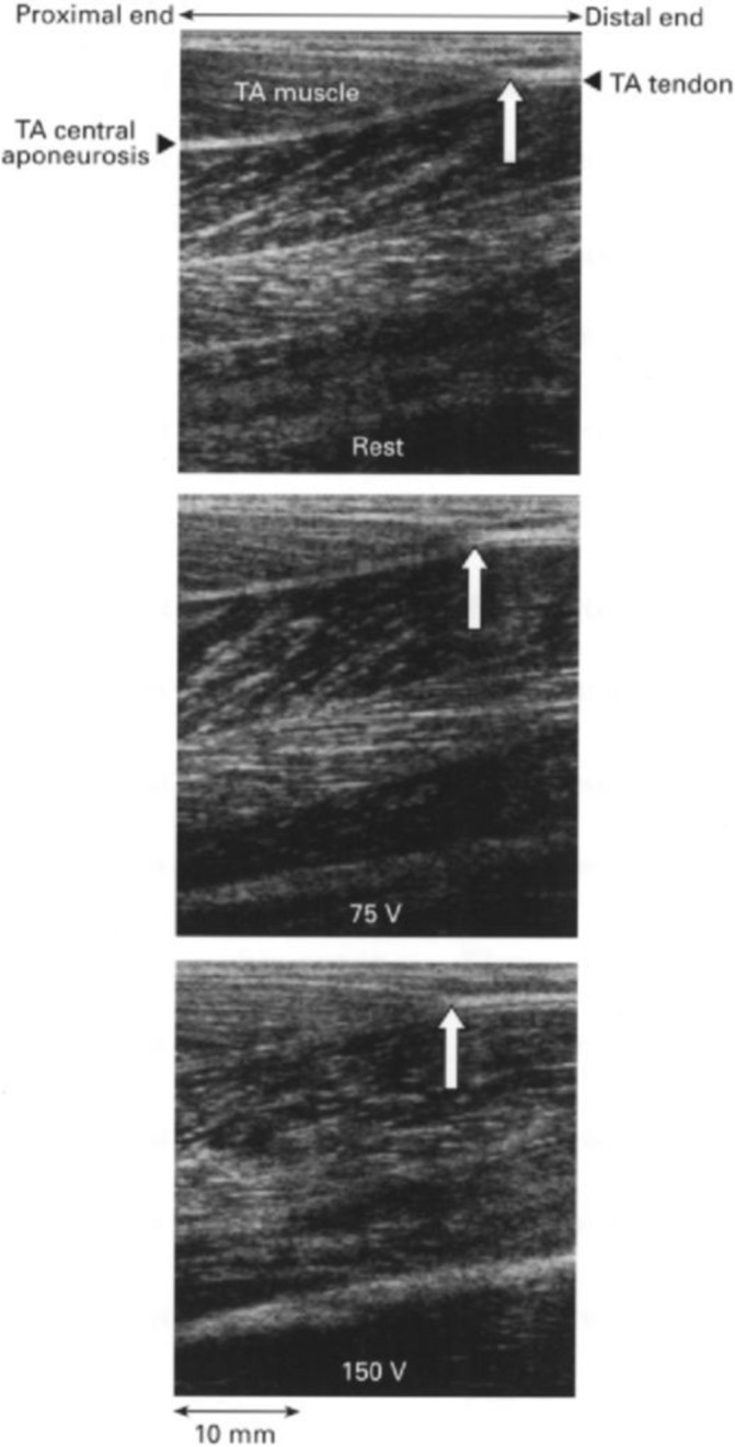
The fascial system builds a three-dimensional continuum of soft, collagen-containing, loose and dense fibrous connective tissue that permeates the body and enables all body systems to operate in an integrated manner. Injuries to the fascial system cause a significant loss of performance in recreational exercise as well as high-performance sports, and could have a potential role in the development and perpetuation of musculoskeletal disorders, including lower back pain. Fascial tissues deserve more detailed attention in the field of sports medicine. A better understanding of their adaptation dynamics to mechanical loading as well as to biochemical conditions promises valuable improvements in terms of injury prevention, athletic performance and sports-related rehabilitation. This consensus statement reflects the state of knowledge regarding the role of fascial tissues in the discipline of sports medicine. It aims to (1) provide an overview of the contemporary state of knowledge regarding the fascial system from the microlevel (molecular and cellular responses) to the macrolevel (mechanical properties), (2) summarise the responses of the fascial system to altered loading (physical exercise), to injury and other physiological challenges including ageing, (3) outline the methods available to study the fascial system, and (4) highlight the contemporary view of interventions that target fascial tissue in sport and exercise medicine. Advancing this field will require a coordinated effort of researchers and clinicians combining mechanobiology, exercise physiology and improved assessment technologies.

Keywords: consensus statement; injury; soft tissue; tendon.


Conflict of interest statement

Competing interests: None declared.

Figures

Figure 1
Figure 1
Components of the fascial system. The fascial system includes large aponeuroses like the first layer of the thoracolumbar fascia (A), but also a myriad of enveloping containers around and within skeletal muscles (B) and most other organs of the body. The internal structure of fascial tissues is dominated by collagen fibres which are embedded in a semiliquid ground substance (C). Images with friendly permission from fascialnet.com (A) and thomas-stephan.com (C).
Figure 2
Figure 2
Transmission electron microscopy reveals the close cell–ECM interaction in human skeletal muscle (musculus vastus lateralis, 25 000× magnification) allowing a bidirectional cell–ECM interaction. Myofilaments (MF) are connected by Z-lines (Z) and costameres (C) to the adjacent basal lamina (BL) and the surrounding reticular lamina (RL). Crossbridging structures (arrows) connect the Z-lines and costameres to the dense part of the basal lamina. The reticular lamina is structured by a network of collagen fibrils (CF) and additional ECM molecules, which have a close connection to the basal lamina allowing bidirectional transmission of mechanical forces. ECM, extracellular matrix.
Figure 3
Figure 3
Factors influencing the mechanical stiffness of fascial tissues and their hypothesised impact. Up arrows symbolise a positive effect (eg, increased cellular contractility increases stiffness), down arrows symbolise a negative effect (eg, increased use of corticosteroids decreases stiffness) and double arrows symbolise an ambiguous association (eg, hyaluronan decreases stiffness if mobilised by mechanical stimuli, but leads to increased stiffness if no stimuli are applied). ECM, extracellular matrix.
Figure 4
Figure 4
Proposed timeline and mechanisms for fascial, adipose and muscle changes in the multifidus muscle after intervertebral disc lesion. Three phases, acute (top), subacute-early chronic (middle) and chronic (bottom), are characterised by different structural and inflammatory changes. IL-1β, interleukin-1β; TNF, tumour necrosis factor.
Figure 5
Figure 5
Tendon displacement measured by B-mode ultrasound. Sonographic images of the human tibialis anterior (TA) muscle at rest (top) and in response to electrical stimulation at 75 V (middle) and 150 V (bottom). The white arrow indicates the TA tendon origin. Notice the proximal shift of the TA tendon origin on electrical stimulation.

References

    1. Adstrum S, Hedley G, Schleip R, et al. . Defining the fascial system. J Bodyw Mov Ther 2017;21:173–7. 10.1016/j.jbmt.2016.11.003 - DOI - PubMed
    1. Ljungqvist A, Schwellnus MP, Bachl N, et al. . International Olympic committee consensus statement: molecular basis of connective tissue and muscle injuries in sport. Clin Sports Med 2008;27:231–9. 10.1016/j.csm.2007.10.007 - DOI - PubMed
    1. Wilke J, Schleip R, Klingler W, et al. . The lumbodorsal fascia as a potential source of low back pain: a narrative review. Biomed Res Int 2017;2017:1–6. 10.1155/2017/5349620 - DOI - PMC - PubMed
    1. Chen B, Ji B, Gao H. Modeling active mechanosensing in cell-matrix interactions. Annu Rev Biophys 2015;44:1–32. 10.1146/annurev-biophys-051013-023102 - DOI - PubMed
    1. Suhr F, Brixius K, Bloch W. Angiogenic and vascular modulation by extracellular matrix cleavage products. Curr Pharm Des 2009;15:389–410. 10.2174/138161209787315756 - DOI - PubMed