. 2019 Mar;21(3):694-704.
doi: 10.1038/s41436-018-0104-7. Epub 2018 Aug 3.

Contribution of noncoding pathogenic variants to RPGRIP1-mediated inherited retinal degeneration

Affiliations

Contribution of noncoding pathogenic variants to RPGRIP1-mediated inherited retinal degeneration

Farzad Jamshidi et al. Genet Med. 2019 Mar.

Abstract

Purpose: With the advent of gene therapies for inherited retinal degenerations (IRDs), genetic diagnostics will have an increasing role in clinical decision-making. Yet the genetic cause of disease cannot be identified using exon-based sequencing for a significant portion of patients. We hypothesized that noncoding pathogenic variants contribute significantly to the genetic causality of IRDs and evaluated patients with single coding pathogenic variants in RPGRIP1 to test this hypothesis.

Methods: IRD families underwent targeted panel sequencing. Unsolved cases were explored by exome and genome sequencing looking for additional pathogenic variants. Candidate pathogenic variants were then validated by Sanger sequencing, quantitative polymerase chain reaction, and in vitro splicing assays in two cell lines analyzed through amplicon sequencing.

Results: Among 1722 families, 3 had biallelic loss-of-function pathogenic variants in RPGRIP1 while 7 had a single disruptive coding pathogenic variants. Exome and genome sequencing revealed potential noncoding pathogenic variants in these 7 families. In 6, the noncoding pathogenic variants were shown to lead to loss of function in vitro.

Conclusion: Noncoding pathogenic variants were identified in 6 of 7 families with single coding pathogenic variants in RPGRIP1. The results suggest that noncoding pathogenic variants contribute significantly to the genetic causality of IRDs and RPGRIP1-mediated IRDs are more common than previously thought.

Keywords: Inherited retinal degeneration; Intronic pathogenic variants; Noncoding pathogenic variants; RPGRIP1; genome sequencing.


Conflict of interest statement

Conflict of interest notification page

The authors declare no conflict of interest related to the work presented in this manuscript.

Figures
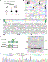
Figure 1.
Figure 1.. Exon 2 duplication in OGI-281.
(a) Pedigree of the family showing deceased parents and the three siblings all of whom were analyzed. (b) qPCR-based copy number results along the first three exons of RPGRIP1. All three siblings have a duplication of exon 2 in an RPGRIP1 allele. Exons 1 and 3 are not affected. The bottom panel shows the locations of RPGRIP1 exons based on the NM_020366 transcript. (c) Integrative genomics viewer (IGV) view of the sequenced whole genome sequencing (WGS) reads where the duplication was discovered for OGI-281–608. The bottom of the figure shows the location of exon 2 of RPGRIP1. The gray thick arrows correspond to expected paired-end reads. The green thick arrows are mapped reads that have aligned abnormally and hint to a mutation. (d) Schematic explanation of the how genomic duplication would lead to the abnormal paired-end reads seen in Figure 1c. The gray region is the area of hypothetical duplication, while the green thick arrows are the paired-end reads that will align abnormally. The top of the figure shows what is actually sequenced in the mutant sample, while the bottom shows how such sequenced reads would map to a reference wild-type (WT) model. The aligned pared-end reads of the mutant will have a greater distance between them and will point away form one another as seen in Figure 1c. The dark and light green hash lines correspond to the aligning sequenced of the paired-end reads. The primers used for Figure 1e are shown as blue and red arrows. Their directionality is indicated relative to the mutant (top) and WT models (bottom). (e) Polymerase chain reaction (PCR) across the predicted duplication breakpoint using primers represented in Figure 1d. Presence of a tandem duplication would yield a product while its absence would lead to no amplification as the primers would be pointing away from each other. The predicted duplication is present in all OGI-281 family members while it is absent in HEK293T cells. The control (Cntrl) PCR on the bottom was done to ensure that larger products could be amplified from all samples thus ensuring DNA fragmentation or quality was not a confounding factor. (f) Sanger sequencing identifying the exact breakpoint (black arrowhead) using OGI-281–608 PCR product from (e). There is a 135bp deletion, upstream of the breakpoint.
Figure 2.
Figure 2.. Exon 1 and 2 duplication in OGI-237 and identification of a novel exon.
(a) Pedigree of OGI-237 showing the segregation of the RPGRIP1 mutations in the family. (b) qPCR of OGI-237 family members, showing the presence of duplication of both exons 1 and 2 in the mother and the proband (523) while the father has a normal copy number across these exons. The bottom panel shows the locations of RPGRIP1 exons based on the NM_020366 transcript. (c) Representation of the predicted WT and mutant alleles with the duplication of exons 1 and 2 (M4). Arrowed lines are introns, tall bars are exons, and short bars indicate untranslated regions (UTR). Further analysis (Figure S3), shows that M4 is a result of tandem duplication with a breakpoint at ~2Kb upstream of exon 1 (black arrow-head). If the transcriptional start site (TSS) is within this 2Kb region, then M4 could lead to normal transcripts given uninterrupted exon 1 and its upstream region. (d) Exploration of the retina transcriptome shows however, that there is an additional novel exon (1n) upstream of the currently annotated exon 1 in the canonical NM_020366 transcript model. Thus the actual TSS is expected to be upstream of exon 1n rather than exon 1. The red bars are indicative of read depth of the transcriptome data. The green and blue arrowed lines at the top indicate split reads between exons. The green are across unannotated exons, while the blue corresponds to annotated exons. There are 550 split reads between exons 1n and 1, further confirming the presence of this novel exon. The NM_020366 canonical transcript model is shown at the bottom. The gray and light blue highlighted areas corresponded to the gray and light blue areas of figure 2e respectively. (e) ATAC-Seq and ChIP-Seq from adult human retina of histone modifications and transcription factor binding at the RPGRIP1 locus as in (d). The light blue shading represents the area directly upstream of the annotated RPGRIP1 transcript. The light gray shading represents a promoter region and newly-discovered retina-specific exon suggested by RNA-Seq, ATAC-Seq and ChIP-Seq. The transcript models of RPGRIP1 are shown on top.
Figure 3.
Figure 3.. Sashimi plots of the amplicon sequencing of splicing assays.
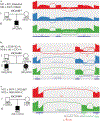
The sashimi plots are generated using amplicon sequencing on RT-PCR products across the indicated regions in HEK293T cells that were transfected with WT (red), positive control (blue), or mutant (green) RPGRIP1 vectors. The annotated NM_020366 exons are indicated on the bottom of each plot in dark blue. (a) In OGI-827, the intronic mutation caused increased intron retention (green), similar to the essential splice site mutation control (blue). (b) In OGI-601, as described in the text, the intronic mutation shifts splice to extend exon 12 (green), as does the essential splice site mutation control (blue). Interestingly, the transcript variant XM_005267881.3 has been reported to include this extention in its exon 1 which is entirely untranslated. Untranslated and translated regions of the transcript models are indicated as thin and thick dark blue lines respectively. (c) In OGI-949, the deep intronic mutation leads to inclusion of a cryptic exon 11’ in the mutant transcript (green). The dashed lines highlight the immediate upstream sequence of the exon 11’ showing the start of the cryptic exon (green) 13bp downstream of the mutation (red). The fourth codon of 11’ is a stop codon (underlined). M11 was a de novo mutation in patient OGI-949–1907, confirmed via Sanger sequencing.
Figure 4.
Figure 4.. Summary of RPGRIP1 mutations and gene model.
Novel findings are indicated in red with non-coding mutations listed below the NM_020366 transcript model and coding mutations listed above. The novel exon 1’ is shown in red. The RPGRIP1 protein and corresponding domains is shown in the bottom.

References

    1. Farrar GJ, Carrigan M, Dockery A, et al. Toward an elucidation of the molecular genetics of inherited retinal degenerations. Hum Mol Genet. August 1 2017;26(R1):R2–R11. - PMC - PubMed
    1. Ellingford JM, Barton S, Bhaskar S, et al. Molecular findings from 537 individuals with inherited retinal disease. J Med Genet. May 11 2016. - PMC - PubMed
    1. Riera M, Navarro R, Ruiz-Nogales S, et al. Whole exome sequencing using Ion Proton system enables reliable genetic diagnosis of inherited retinal dystrophies. Sci Rep. February 9 2017;7:42078. - PMC - PubMed
    1. Bujakowska KM, Fernandez-Godino R, Place E, et al. Copy-number variation is an important contributor to the genetic causality of inherited retinal degenerations. Genetics in medicine : official journal of the American College of Medical Genetics. June 2017;19(6):643–651. - PMC - PubMed
    1. Liquori A, Vache C, Baux D, et al. Whole USH2A Gene Sequencing Identifies Several New Deep Intronic Mutations. Human mutation. February 2016;37(2):184–193. - PubMed

Publication types