Review
. 2018 Jul;84(Suppl 1):46-56.
doi: 10.1038/s41390-018-0077-x.

Application of NPE in the assessment of a patent ductus arteriosus

Collaborators, Affiliations
Review

Application of NPE in the assessment of a patent ductus arteriosus

David van Laere et al. Pediatr Res. 2018 Jul.

Abstract

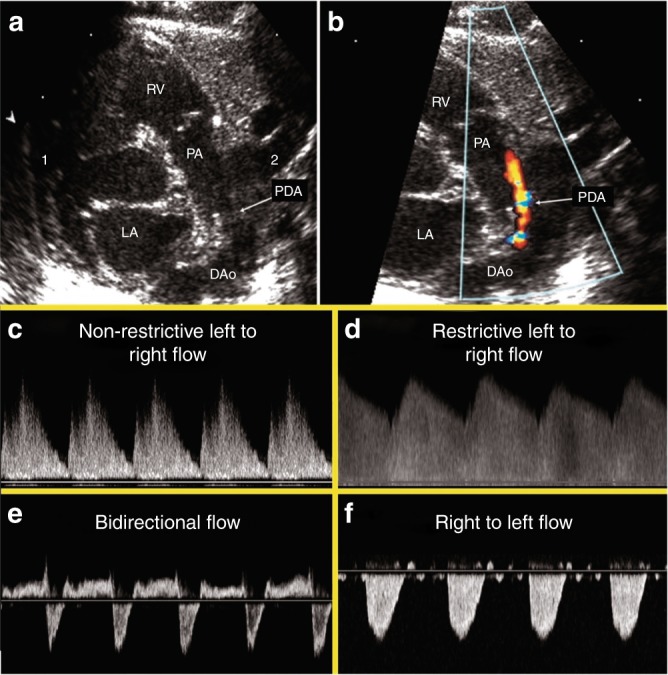
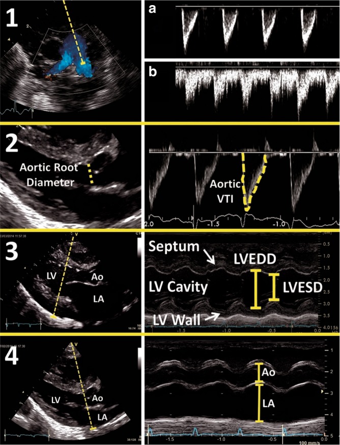
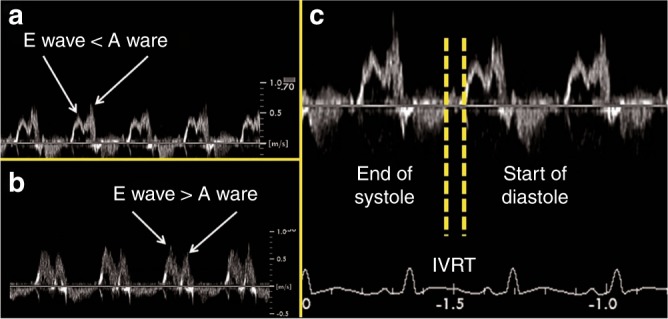
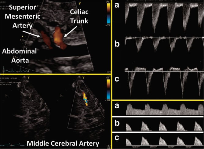
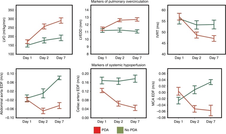
In many preterm infants, the ductus arteriosus remains patent beyond the first few days of life. This prolonged patency is associated with numerous adverse outcomes, but the extent to which these adverse outcomes are attributable to the hemodynamic consequences of ductal patency, if at all, has not been established. Different treatment strategies have failed to improve short-term outcomes, with a paucity of data on the correct diagnostic and pathophysiological assessment of the patent ductus arteriosus (PDA) in association with long-term outcomes. Echocardiography is the selected method of choice for detecting a PDA, assessing the impact on the preterm circulation and monitoring treatment response. PDA in a preterm infant can result in pulmonary overcirculation and systemic hypoperfusion, Therefore, echocardiographic assessment should include evaluation of PDA characteristics, indices of pulmonary overcirculation with left heart loading conditions, and indices of systemic hypoperfusion. In this review, we provide an evidence-based overview of the current and emerging ultrasound measurements available to identify and monitor a PDA in the preterm infant. We offer indications and limitations for using Neonatologist Performed Echocardiography to optimize the management of a neonate with a PDA.


Conflict of interest statement

A.E.K. is in receipt of an Irish Health Research Board Clinical Trials Network Grant (HRB CTN 2014-10) and an EU FP7/2007-2013 grant (agreement no. 260777, The HIP Trial). A.G. owned equity in Neonatal Echo Skills and has received grant support from the American Heart Association. E.D. received lecture fees and consulting fees from Chiesi Pharmaceutical. E.N. received grant support from Research Council of Norway and Vestfold Hospital Trust. K.B. received lecture fees from Chiesi Pharmaceutical. M.B. holds a patent “Thermal shield for the newborn baby”. S.R. received lecture fees for Phillips Ultrasound and GE Ultrasound. W.P.B. has received grant support from The Netherlands Organization for Health and Development (ZonMw; grant number 843002608). Z.M. has received lecture fees from Chiesi Pharmaceutical. The remaining authors declared no competing interests.

Figures

Fig. 1
Fig. 1
Chest radiograph in a ventilated preterm infant with a large PDA. Note the large heart shadow and the increased lung markings representing pulmonary overcirculation
Fig. 2
Fig. 2
PDA 2D, color Doppler image and Doppler flow patterns. The top panels demonstrate the PDA in 2D (a) and color Doppler (b). c Pulsatile or non-restrictive pattern: characterized by a left to right (LtR) shunt with an arterial waveform and high peak systolic velocity: end-diastolic velocity ratio. d Restrictive pattern: characterized by high systolic and diastolic velocity, and low peak systolic velocity: end-diastolic velocity ratio. e Bidirectional pattern: elevated pulmonary pressures equal to or near systemic. f Right to left (RtL) flow: supra-systemic pulmonary pressures
Fig. 3
Fig. 3
Assessment of left heart volume loading. (1) Measurement of diastolic flow in the left pulmonary artery. a Normal situation without ductal left-to-right shunting. b Illustrates forward diastolic flow in the presence of significant left-to-right ductal flow. (2) Measurement of LVO: increased LVO in the setting of a PDA indicated increased pulmonary venous return. (3) Measurement of LV diameter in diastole: increased LV diameter is another surrogate marker for increased LV end-diastolic volume. (4) LA:Ao ratio: atrial enlargement can be indexed to a relatively fixed aortic root diameter to further estimate in degree of increased LA volume
Fig. 4
Fig. 4
Assessment of left heart pressure loading. Transmitral LV filling in normal term infants is characterized by a predominance of early diastolic (“E”) filling, with limited late LV filling occurring during atrial contraction (“A”), resulting in an E:A ratio >1. a Healthy preterm infants without a PDA have intrinsically decreased LV diastolic function, relying more on late atrial filling, and E:A ratio <1. b and c Preterm infants with a large PDA have increased left atrial pressure which results in earlier mitral valve opening and drives early passive filling, resulting in shortened isovolumic relaxation time (<40 ms) and a “pseudonormalized” E:A ratio >1
Fig. 5
Fig. 5
Assessment of diastolic flow in a post-ductal artery. Measurement of pulsed wave Doppler pattern in the celiac trunk, the abdominal aorta, and the middle cerebral can highlight the effect of left-to-right shunting across the PDA. In the top Doppler panel, three abdominal aortic Doppler wave forms are illustrated demonstrating normal forward diastolic flow (a), absent diastolic flow (b), and revered diastolic flow (c). A similar pattern can be seen in the lower Doppler panel which is representative of celiac and middle cerebral arteries
Fig. 6
Fig. 6
Patterns of echocardiography markers in infants with and without a PDA over the first week of age. Divergence in echocardiography parameters becomes apparent within the first 48 h following birth. Data represent means and standard error (data adapted from EL-Khuffash et al.). LVO left ventricular output, LVEDD left ventricular end-diastolic diameter, IVRT isovolumic relaxation time, EDF end-diastolic velocity, MCA middle cerebral artery

Comment in

References

    1. Reese J, Shelton EL, Slaughter JC, McNamara PJ. Prophylactic indomethacin revisited. J. Pediatr. 2017;186:11–14. doi: 10.1016/j.jpeds.2017.03.036. - DOI - PMC - PubMed
    1. Weber SC, et al. Natural history of patent ductus arteriosus in very low birth weight infants after discharge. J. Pediatr. 2015;167:1149–1151. doi: 10.1016/j.jpeds.2015.06.032. - DOI - PubMed
    1. Levy PT, et al. Maturational patterns of systolic ventricular deformation mechanics by two-dimensional speckle-tracking echocardiography in preterm infants over the first year of age. J. Am. Soc. Echocardiogr. 2017;30:685–698. doi: 10.1016/j.echo.2017.03.003. - DOI - PMC - PubMed
    1. Benitz WE, Committee On F, Newborn. Patent ductus arteriosus in preterm infants. Pediatrics. 2016;137:1–6. doi: 10.1542/peds.2015-3730. - DOI - PubMed
    1. Ramos FG, Rosenfeld CR, Roy L, Koch J, Ramaciotti C. Echocardiographic predictors of symptomatic patent ductus arteriosus in extremely-low-birth-weight preterm neonates. J. Perinatol. 2010;30:535–539. doi: 10.1038/jp.2010.14. - DOI - PubMed

Publication types

MeSH terms