Review
. 2018 Jul;84(Suppl 1):1-12.
doi: 10.1038/s41390-018-0076-y.

Introduction to neonatologist-performed echocardiography

Collaborators, Affiliations
Review

Introduction to neonatologist-performed echocardiography

Alan M Groves et al. Pediatr Res. 2018 Jul.

Abstract

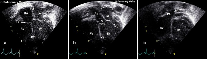
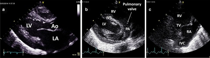
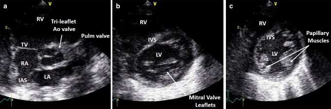
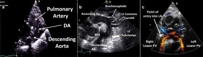
Cardiac ultrasound techniques are increasingly used in the neonatal intensive care unit to guide cardiorespiratory care of the sick newborn. This is the first in a series of eight review articles discussing the current status of "neonatologist-performed echocardiography" (NPE). The aim of this introductory review is to discuss four key elements of NPE. Indications for scanning are summarized to give the neonatologist with echocardiography skills a clear scope of practice. The fundamental physics of ultrasound are explained to allow for image optimization and avoid erroneous conclusions from artifacts. To ensure patient safety during echocardiography recommendations are given to prevent cardiorespiratory instability, hypothermia, infection, and skin lesions. A structured approach to echocardiography, with the same standard views acquired in the same sequence at each scan, is suggested in order to ensure that the neonatologist confirms normal structural anatomy or acquires the necessary images for a pediatric cardiologist to do so when reviewing the scan.


Conflict of interest statement

A.E.-K. is in receipt of an Irish Health Research Board Clinical Trials Network Grant (HRB CTN 2014-10) and an EU FP7/2007-2013 grant (agreement no. 260777, The HIP Trial). A.M.G. owned equity in Neonatal Echo Skills and has received grant support from the American Heart Association. D.V.L. is in receipt of an EU FP7/2007-2013 (agreement no 260777 the HIP trial). E.D. received lecture fees and consulting fees from Chiesi Pharmaceutical. E.N. received grant support from Research Council of Norway and Vestfold Hospital Trust. K.B. received lecture fees from Chiesi Pharmaceutical. M.B. holds a patent, “Thermal shield for the newborn baby. S.G. received grant support from National Institute of Health Research, Health Technology Assessment (11/92/15), UK. S.R.R. received lecture fees for Phillips Ultrasound and GE Ultrasound. W.P.B. has received grant support from The Netherlands Organization for Health and Development (ZonMw; grant number 843002622 and 843002608). Z.M. has received lecture fees from Chiesi Pharmaceutical. The other authors declare no competing interests.

Figures

Fig. 1
Fig. 1
Parameters of a sound wave
Fig. 2
Fig. 2
Pulse duration and pulse repetition period. Adapted from Rovner A. The principle of ultrasound—ECHOpedia 2017. Available from: http://www.echopedia.org/wiki/The_principle_of_ultrasound
Fig. 3
Fig. 3
Ultrasound and tissue interactions. Adapted from Textbook of Clinical Echocardiography, 5th Edition, Catherine Otto, “Principles of Echocardiographic Image Acquisition and Doppler Analysis,” Fig. 1.4, page 5, Copyright (2013), with permission from Elsevier
Fig. 4
Fig. 4
Axial (left panel) and lateral (right panel) resolution. See text for details. Adapted from Rovner A. The principle of ultrasound—ECHOpedia 2017. Available from: http://www.echopedia.org/wiki/The_principle_of_ultrasound
Fig. 5
Fig. 5
Arch view in a newborn showing reverberation and sidelobe artifacts
Fig. 6
Fig. 6
A subcostal situs view demonstrating an echogenic focus in the liver and shadow artifact (a) and a subcostal parasagittal view of the liver and inferior vena cava (IVC) demonstrating a mirror image against the diaphragm (b)
Fig. 7
Fig. 7
An example of beam width artifact in the parasternal short axis view at the level of the papillary muscle
Fig. 8
Fig. 8
Doppler shift and angle of insonation (see text for details). Adapted from Rovner A. The principle of ultrasound—ECHOpedia 2017. Available from: http://www.echopedia.org/wiki/The_principle_of_ultrasound. f0 transmitted frequency, fR received frequency; ∆f Doppler shift, ν blood velocity, cos Ø cosine of insonation angle, c ultrasound velocity in blood
Fig. 9
Fig. 9
Continuous-wave (CW) and pulsed-wave (PW) Doppler. Signal sampling and aliasing (see text for details). Adapted from Rovner A. The principle of ultrasound— ECHOpedia 2017. Available from: http://www.echopedia.org/wiki/The_principle_of_ultrasound
Fig. 10
Fig. 10
Transverse subcostal view (a), right parasagittal subcostal view (b), and left parasagittal subcostal view (c). IVC inferior vena cava, RA right atrium
Fig. 11
Fig. 11
Subcostal atrial view 2D (a) and color Doppler showing left-to-right interatrial shunting (b)
Fig. 12
Fig. 12
Subcostal view showing crossing great arteries; aorta (a) and pulmonary artery (b). IVS inter-ventricular septum, LV left ventricle, PA pulmonary artery, RV right ventricle, RA right atrium, LA left atrium
Fig. 13
Fig. 13
Apical four-chamber view (a), LV outflow tract (b), and RV outflow tract (c). IAS interatrial septum, IVS inter-ventricular septum, Ao aorta, LA left atrium, LV left ventricle, MV mitral valve, RA right atrium, RV right ventricle, TV tricuspid valve, LPA left pulmonary artery
Fig. 14
Fig. 14
Parasternal long axis view of the LV (a), pulmonary artery (b), and tricuspid valve (c). Ao aorta, LV left ventricle, LA left atrium, IVS inter-ventricular septum, RV right ventricle, TV tricuspid valve, IVC inferior vena cava
Fig. 15
Fig. 15
Parasternal short axis view at the level of the aorta (a), mitral valve (b), and papillary muscles (c). Ao aorta, LV left ventricle, LA left atrium, IVS inter-ventricular septum, IAS interatrial septum, RV right ventricle, TV tricuspid valve, IVC inferior vena cava
Fig. 16
Fig. 16
Ductal view (a), arch view (b), and (c). Ao aorta, DA ductus arteriosus, LA left atrium, Lt left, PV pulmonary vein, RPA right pulmonary artery

Comment in

References

    1. Skinner JR. Echocardiography on the neonatal unit: a job for the neonatologist or the cardiologist? Arch. Dis. Child. 1998;78:401–402. doi: 10.1136/adc.78.5.401. - DOI - PMC - PubMed
    1. El-Khuffash AF, McNamara PJ. Neonatologist-performed functional echocardiography in the neonatal intensive care unit. Semin. Fetal Neonatal Med. 2011;16:50–60. doi: 10.1016/j.siny.2010.05.001. - DOI - PubMed
    1. Evans N, et al. Point-of-care ultrasound in the neonatal intensive care unit: international perspectives. Semin. Fetal Neonatal Med. 2011;16:61–68. doi: 10.1016/j.siny.2010.06.005. - DOI - PubMed
    1. Mertens L, et al. Targeted Neonatal Echocardiography in the Neonatal Intensive Care Unit: practice guidelines and recommendations for training. Writing Group of the American Society of Echocardiography (ASE) in collaboration with the European Association of Echocardiography (EAE) and the Association for European Pediatric Cardiologists (AEPC) J. Am. Soc. Echocardiogr. 2011;24:1057–1078. doi: 10.1016/j.echo.2011.07.014. - DOI - PubMed
    1. Singh Y, et al. Expert consensus statement “Neonatologist-performed Echocardiography (NoPE)”—training and accreditation in UK. Eur. J. Pediatr. 2015;175:281–287. doi: 10.1007/s00431-015-2633-2. - DOI - PubMed

Publication types

MeSH terms