Case Reports
. 2018 Jul 8:2018:2037638.
doi: 10.1155/2018/2037638. eCollection 2018.

Treatment of a Neglected Patellar Tendon Rupture with a Modified Surgical Technique: Ipsilateral Semitendinosus Autograft Reconstruction with Suture Tape Augmentation

Affiliations
Case Reports

Treatment of a Neglected Patellar Tendon Rupture with a Modified Surgical Technique: Ipsilateral Semitendinosus Autograft Reconstruction with Suture Tape Augmentation

Sanjum P Samagh et al. Case Rep Orthop. .

Abstract

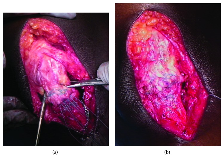
Patellar tendon ruptures are rare, but debilitating injuries are typically seen in young active males in the third and fourth decades of life. They can occur as a single acute injury or from repetitive microtrauma weakening the tendon. Patients typically present complaining of knee pain, swelling, and an inability to perform a straight leg raise. Most conventionally, these injuries are classified as acute (less than two weeks) or chronic (greater than two weeks) based upon the timing of presentation. In patients with patellar tendon ruptures and inability to perform a straight leg raise, patellar tendon repair is most often recommended. A subset of patients with chronic patellar tendon ruptures, however, presents several months after their initial injuries. These neglected patella tendon ruptures present a particularly challenging clinical scenario in which primary repair is often difficult or not possible. This case report describes a modification to an existing surgical technique for reconstructing the patellar tendon using an ipsilateral semitendinosus tendon autograft with suture tape augmentation.


Figures

Figure 1
Figure 1
(a) The peritenon is identified superiorly, and peritendonous flaps are elevated to reveal the chronic, scarred patellar tendon rupture (approximately 3.5 cm in the midsubstance of the tendon). (b) The chronically scarred portion of the tendon is then debrided along with the medial and lateral retinacula. A Cobb elevator is used to release the superficial and deep aspects of the quadriceps tendon from the surrounding tissues.
Figure 2
Figure 2
(a) A four-stranded end-to-end repair is then undertaken using a #5 FiberWire (Arthrex, Naples, FL) in a Krackow fashion. (b) To secure the repair, a #2 FiberWire (Arthrex, Naples, FL) is used in a running-locking fashion to imbricate the elongated medial and lateral retinacula and oversew the tendon repair.
Figure 3
Figure 3
(a) The semitendinosus autograft is passed through the bone tunnel from medial to lateral and courses along the lateral aspect of the native patellar tendon. Small rents are created in the medial and lateral retinacula at the level of the superior pole of the patella, and the graft is woven transversely through the distal quadriceps tendon from lateral to medial. The graft is then brought down along the medial border of the native patellar tendon and passed through the bone tunnel from lateral to medial. The free ends cross in opposite directions within the tibial bone tunnel and are secured within the tibial tunnel using two interference screws (Tenodesis Screw, BioComposite, 6.25 × 15 mm; Arthrex, Naples, FL)—one from medial to lateral and one from lateral to medial. (b) An InternalBrace (Arthrex, Naples, FL) is used to augment and protect the reconstruction. Two 4.75 × 15 mm SwiveLock BioComposite suture anchors (Arthrex, Naples, FL) are placed into the distal pole of the patella securing the midpoint of one 2 mm FiberTape suture tape (Arthrex, Naples, FL) each. The free limbs of the FiberTape (Arthrex, Naples, FL) cross the repair site—one limb straight inferior and the other in an “X” fashion—and are then secured to the tibial tubercle using two 4.75 × 15 mm SwiveLock BioComposite suture anchors (Arthrex, Naples, FL).

References

    1. Siwek C. W., Rao J. P. Ruptures of the extensor mechanism of the knee joint. The Journal of Bone & Joint Surgery. 1981;63(6):932–937. doi: 10.2106/00004623-198163060-00010. - DOI - PubMed
    1. Zernicke R. F., Garhammer J., Jobe F. W. Human patellar-tendon rupture. The Journal of Bone & Joint Surgery. 1977;59(2):179–183. doi: 10.2106/00004623-197759020-00007. - DOI - PubMed
    1. Kelly D. W., Carter V. S., Jobe F. W., Kerlan R. K. Patellar and quadriceps tendon ruptures—jumper's knee. The American Journal of Sports Medicine. 1984;12(5):375–380. doi: 10.1177/036354658401200508. - DOI - PubMed
    1. Hsu K. Y., Wang K. C., Ho W. P., Hsu R. W. Traumatic patellar tendon ruptures: a follow-up study of primary repair and a neutralization wire. The Journal of Trauma: Injury, Infection, and Critical Care. 1994;36(5):658–660. doi: 10.1097/00005373-199405000-00010. - DOI - PubMed
    1. Lamberti A., Balato G., Summa P. P., Rajgopal A., Vasdev A., Baldini A. Surgical options for chronic patellar tendon rupture in total knee arthroplasty. Knee Surgery, Sports Traumatology, Arthroscopy. 2018;26(5):1429–1435. doi: 10.1007/s00167-016-4370-0. - DOI - PubMed

Publication types

LinkOut - more resources