. 2018 Aug 1;59(10):3808-3815.
doi: 10.1167/iovs.18-24118.

Murine Retinal Citrullination Declines With Age and is Mainly Dependent on Peptidyl Arginine Deiminase 4 (PAD4)

Affiliations

Murine Retinal Citrullination Declines With Age and is Mainly Dependent on Peptidyl Arginine Deiminase 4 (PAD4)

T J Hollingsworth et al. Invest Ophthalmol Vis Sci. .

Abstract

Purpose: Citrullination is a post-translational modification (PTM) that serves many normal physiological functions. Studies have shown that this PTM-along with expression of the catalyzing enzymes, peptidyl arginine deiminases (PADs)-are increased in autoimmune and age-related pathologies. PAD2 retinal expression has been previously documented in rat and human. Herein, we report on the expression levels and patterns of PAD2, PAD4, and retinal citrullination in the murine retina with age.

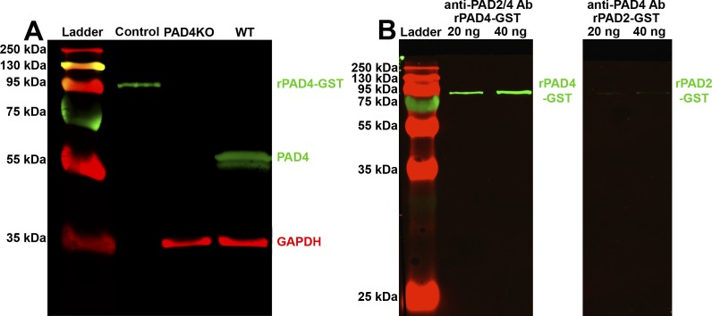
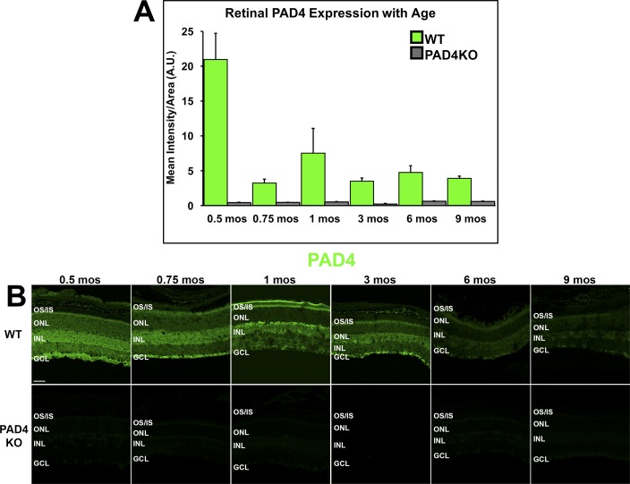
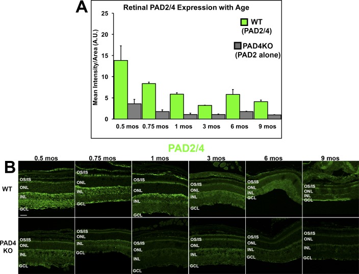
Methods: Wild-type (WT) and Pad4-/- (PAD4KO) mice ages 0.5, 0.75, 1, 3, 6, and 9 months were investigated after euthanasia and eye enucleation. Retinal lysates from 3-month-old mice were probed for PAD4 by western blot. Whole eyes were fixed, cryosectioned, and probed using an anti-PAD2/4 antibody (Ab), a specific anti-PAD4 Ab, and F95 anti-citrullinated peptide Ab. Fluorescent intensities were quantified with ImageJ.

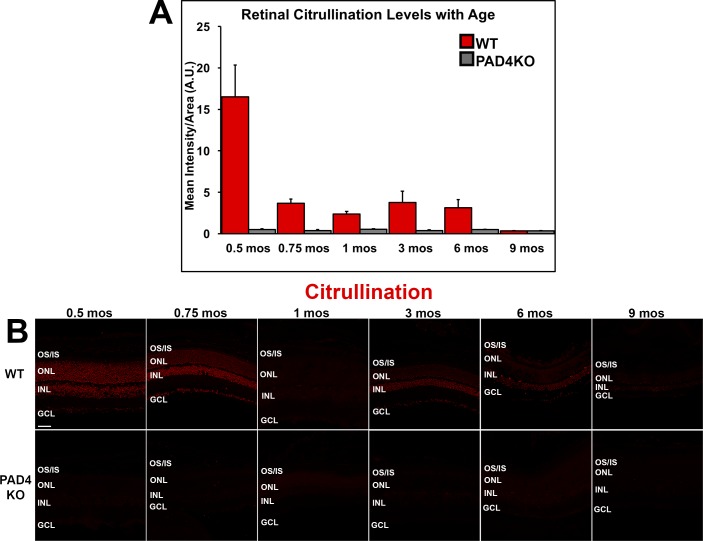
Results: WT retinas show different levels of PAD4 expression in distinct retinal layers, with the most intense labeling in inner retinal layers, while PAD4KO mice lacked retinal PAD4. Using a nonspecific anti-PAD2/4 Ab, PAD reactivity observed in PAD4KO mice was attributed to PAD2. In WT, both PAD2 and PAD4 expression levels decrease significantly with age while low-level residual PAD2 expression was seen in PAD4KO mice. Citrullination levels in WT retinas paralleled PAD4 expression, with PAD4KO mice exhibiting consistently minimal citrullination.

Conclusions: Both PAD2 and PAD4 expression and citrullination decrease with age in the murine retina. However, in the absence of PAD4, retinal citrullination is nearly abolished, indicating that PAD4 is a main effector for retinal citrullination under physiological conditions.


Figures

Figure 1
Figure 1
Western blot analysis of PAD4 expression in mouse retinal lysate and testing for antibody cross-reactivity. (A) Retinal lysates from WT and PAD4KO mice were electrophoresed and probed for PAD4 (green). GAPDH (red) served as a loading control. Control, 20 ng PAD4-GST fusion protein (positive control). At 3 months, WT mice exhibit a strong band at 65 kDa, indicative of PAD4, while PAD4KO animals exhibit no band, indicative of loss of PAD4. (B) 20 ng and 40 ng of rPAD4-GST (lanes 2 and 3) and rPAD2-GST (lanes 4 and 5) were electrophoresed and probed for PAD2 and PAD4 (both green), respectively. The anti-PAD2/4 antibody bound to PAD4 with a high affinity while the anti-PAD4 antibody bound negligibly to PAD2 protein.
Figure 2
Figure 2
IHC analysis of PAD4 localization and expression levels in the mouse retina. (A) PAD4 expression levels were quantified using densitometric analysis of fluorescent intensities across the neural retina using ImageJ and plotted as mean intensity divided by area as a function of age. Data are expressed as the mean ± SEM (error bars, n = 3). A steep decline in immunoreactivity was seen in WT mice between 0.5 and 0.75 months (14 vs. 21 days) of age, with subsequent peaks at 1 month and between 3 and 6 months. (B) Immunolabeling of PAD4 (green) in WT and PAD4KO mouse retinas at serial time points. GCL, ganglion cell layer; INL, inner nuclear layer; ONL, outer nuclear layer; OS/IS, photoreceptor outer segments/inner segments. Scale bar: 50 μm.
Figure 3
Figure 3
IHC localization and expression levels of total PAD (WT) and PAD2 (PAD4KO) in the mouse retina. (A) Expression levels of total PAD (WT) and residual PAD2 (PAD4KO) were quantified using densitometric analysis of fluorescent intensities across the neural retina using ImageJ and plotted as mean intensity divided by area as a function of age. Data are expressed as the mean ± SEM (error bars, n = 3). A decline in both total PAD immunoreactivity and residual PAD2 reactivity over time was seen in both WT mice and PAD4KO mice, respectively. (B) Immunolabeling (in green) attributable to both PAD2 and PAD4 (total PAD reactivity) in WT mice and residual PAD2 reactivity seen in PAD4KO mouse retinas at serial time points. GCL, ganglion cell layer; INL, inner nuclear layer; ONL, outer nuclear layer; OS/IS, photoreceptor outer segments/inner segments. Scale bar: 50 μm.
Figure 4
Figure 4
Immunohistochemical analysis of citrullinated peptide localization and levels in the mouse retina. (A) Citrullinated peptide expression levels were quantified using densitometric analysis of fluorescent intensities across the neural retina using ImageJ and plotted as mean intensity divided by area as a function of age. Data are expressed as the mean ± SEM (error bars, n = 6). Reactivity declined over time, reaching negligible levels by 9 months under physiological conditions. (B) Immunolabeling of citrullinated peptides (red) in WT and PAD4KO mouse retinas at increasing ages. The pattern of expression of citrullinated peptides over time mirrored closest the dynamics of PAD4 expression. GCL, ganglion cell layer; INL, inner nuclear layer; ONL, outer nuclear layer; OS/IS, photoreceptor outer segments/inner segments. Scale bar: 50 μm.

References

    1. Vossenaar ER, Zendman AJ, van Venrooij WJ, Pruijn GJ. PAD. a growing family of citrullinating enzymes: genes, features and involvement in disease. Bioessays. 2003;25:1106–1118. - PubMed
    1. Witalison EE, Thompson PR, Hofseth LJ. Protein arginine deiminases and associated citrullination: physiological functions and diseases associated with dysregulation. Curr Drug Targets. 2015;16:700–710. - PMC - PubMed
    1. Acharya NK, Nagele EP, Han M., et al. Neuronal PAD4 expression and protein citrullination: possible role in production of autoantibodies associated with neurodegenerative disease. J Autoimmun. 2012;38:369–380. - PubMed
    1. Calabrese R., Zampieri M., Mechelli R., et al. Methylation-dependent PAD2 upregulation in multiple sclerosis peripheral blood. Mult Scler. 2012;18:299–304. - PubMed
    1. Moscarello MA, Mastronardi FG, Wood DD. The role of citrullinated proteins suggests a novel mechanism in the pathogenesis of multiple sclerosis. Neurochem Res. 2007;32:251–256. - PMC - PubMed

Publication types

Substances