Review
. 2019 Sep;138(8-9):917-936.
doi: 10.1007/s00439-018-1918-8. Epub 2018 Aug 2.

The hedgehog pathway and ocular developmental anomalies

Affiliations
Review

The hedgehog pathway and ocular developmental anomalies

Florencia Cavodeassi et al. Hum Genet. 2019 Sep.

Abstract

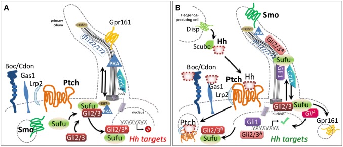
Mutations in effectors of the hedgehog signaling pathway are responsible for a wide variety of ocular developmental anomalies. These range from massive malformations of the brain and ocular primordia, not always compatible with postnatal life, to subtle but damaging functional effects on specific eye components. This review will concentrate on the effects and effectors of the major vertebrate hedgehog ligand for eye and brain formation, Sonic hedgehog (SHH), in tissues that constitute the eye directly and also in those tissues that exert indirect influence on eye formation. After a brief overview of human eye development, the many roles of the SHH signaling pathway during both early and later morphogenetic processes in the brain and then eye and periocular primordia will be evoked. Some of the unique molecular biology of this pathway in vertebrates, particularly ciliary signal transduction, will also be broached within this developmental cellular context.


Figures

Fig. 1
Fig. 1
a Before binding by a Hedgehog family ligand (most often Shh, but also Ihh or Dhh), Ptch1 and presumably Ptch2 maintain Smo sequestered, by a currently unknown mechanism. Under these circumstances, both anterograde and retrograde transport in functionally intact primary cilia is necessary to allow PKA-dependent cleavage of the Gli3 transcription factor to its repressor form and its shuttling from the base of the cilium to target DNA, accompanied by Sufu. The same mechanism may govern Gli2 processing. b Hedgehog proteins are post-translationally modified; the secretion and multimeric conformation of Shh is organized by the Disp transmembrane receptor in signaling cells, accompanied by Scube. Binding of Hh to Ptch receptors leads to internalization and degradation, and relocalization of Smo to the primary ciliary membrane, which changes lipid composition. Other receptors include Boc, Cdon and Gas1, which may refine signal integration. Lrp1 is a non-specific but necessary co-receptor. Hh binding leads to ciliary clearance of the G-protein coupled negative regulatory receptor Gpr161, reduction in PKA production, and accompaniment by Sufu of immature Gli2/3 proteins along the ciliary microtubules for their requisite processing to activating forms. Gli1 is transcribed as a Shh target and positively reinforces this cycle, while Ptch1 itself is another target whose increased transcription sharpens thresholds and raises cellular stakes for survival, as continued Hh stimulation becomes necessary for the target cell. The activity of cell cycle-related kinase (Ccrk) is necessary for ciliary integrity and acts between Smo and Gli2 in effecting readout of Shh levels within the optic primordium
Fig. 2
Fig. 2
Ptch1 mRNA expression in the adult mouse retina (as reported in Chassaing et al. 2016). Transcripts in bluish-purple are present within all cell bodies of the retina. From external to internal: scl sclera, ch choroid plexus, heavily invested with melanocytes, RPE retinal pigmented epithelium, ES external segment of photoreceptors—both rods and cones in contact with the RPE, elm the external limiting “membrane” (aligned tight junctions of photoreceptors and Müller cells), EGL external granular layer, composed of photoreceptor soma, EPL external plexiform layer, which contains the dendrites of bipolar and horizontal interneurons that integrate photoreceptor signals, IGL internal granular layer, with the cell bodies of the bipolar, horizontal and amacrine cells, IPL internal plexiform layer, with the axons of bipolar cells connected to amacrine cells and retinal ganglion cell (RGC) dendrites, RGC cell body layer, NL nerve fiber layer of RGC axons converging on the optic nerve, ILM inner limiting membrane, vitr vitreous

References

    1. Acampora D, Mazan S, Lallemand Y, et al. Forebrain and midbrain regions are deleted in Otx2/ mutants due to a defective anterior neuroectoderm specification during gastrulation. Development. 1995;121:3279–3290. - PubMed
    1. Aguiar DP, Sghari S, Creuzet S. The facial neural crest controls fore- and midbrain patterning by regulating Foxg1 expression through Smad1 activity. Development. 2014;141:2494–2505. - PubMed
    1. Ahlgren SC, Bronner-Fraser M. Inhibition of sonic hedgehog signaling in vivo results in craniofacial neural crest cell death. Curr Biol. 1999;9:1304–1314. - PubMed
    1. Alsaif HS, Khan AO, Patel N, et al. Congenital glaucoma and CYP1B1: an old story revisited. Hum Genet. 2018 - PubMed
    1. Aoto K, Trainor PA. Co-ordinated brain and craniofacial development depend upon Patched1/XIAP regulation of cell survival. Hum Mol Genet. 2015;24:698–713. - PMC - PubMed

Substances

LinkOut - more resources