. 2018 Dec;256(12):2449-2456.
doi: 10.1007/s00417-018-4081-y. Epub 2018 Aug 2.

CD163+ macrophages infiltrate axon bundles of postmortem optic nerves with glaucoma

Affiliations

CD163+ macrophages infiltrate axon bundles of postmortem optic nerves with glaucoma

Milica A Margeta et al. Graefes Arch Clin Exp Ophthalmol. 2018 Dec.

Abstract

Purpose: Prior research in animal models has shown that macrophages and microglia play an important role in pathogenesis of glaucoma, but the phenotype and distribution of macrophages in human glaucomatous tissue have not been sufficiently characterized.

Methods: We analyzed H&E, CD68-, and CD163-immunostained slides from 25 formaldehyde-fixed, paraffin-embedded autopsy eyes: 12 control eyes and 13 eyes with glaucoma. The diagnosis of glaucoma was made based on a history of glaucoma as reported in the medical record and histological changes characteristic of glaucoma. Glaucoma cases and controls were matched in terms of age, sex, and race.

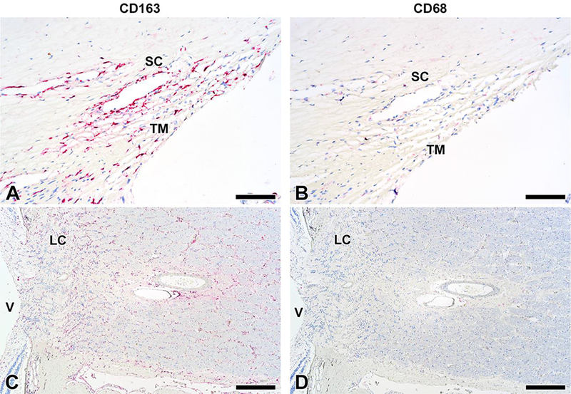
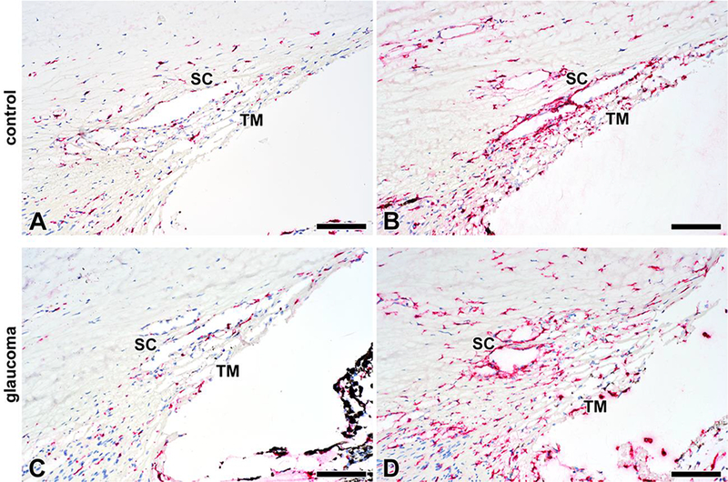
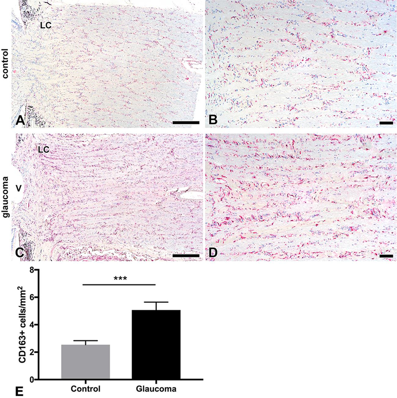
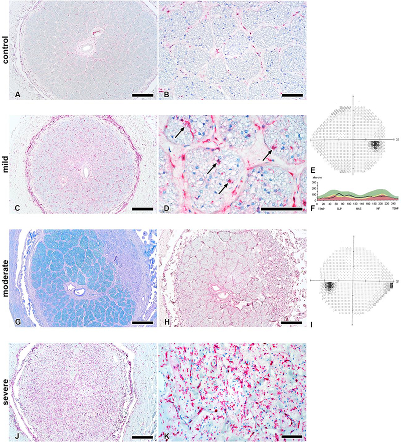
Results: Qualitative analysis of the conventional outflow pathway and the optic nerve revealed that all eyes contained CD163+ cells but a negligible number of CD68+ cells. CD163+ macrophages infiltrated the trabecular meshwork and surrounded Schlemm's canal of normal eyes and eyes with glaucoma, but the pattern was variable and qualitatively similar between groups. In optic nerves of control eyes, CD163+ macrophages were present at low levels and restricted to septa between axon bundles. In glaucomatous optic nerves, the number of CD163+ cells was increased both qualitatively and quantitatively (glaucoma 5.1 ± 0.6 CD163+ cells/mm2, control 2.5 ± 0.3 CD163+ cells/mm2, p < 0.001), with CD163+ cells infiltrating axon bundles in cases of both mild and severe diseases.

Conclusions: The increase in CD163+ cell number in eyes with mild and severe glaucoma is the first demonstration of macrophage infiltration in glaucomatous human optic nerves. This finding supports a role for macrophages in glaucoma pathogenesis and progression.

Keywords: Glaucoma; Macrophages; Microglia; Neurodegeneration; Neuroprotection; Optic nerve.


Conflict of interest statement

Conflict of Interest: All authors certify that they have no affiliations with or involvement in any organization or entity with any financial interest (such as honoraria; educational grants; participation in speakers’ bureaus; membership, employment, consultancies, stock ownership, or other equity interest; and expert testimony or patent-licensing arrangements), or non-financial interest (such as personal or professional relationships, affiliations, knowledge or beliefs) in the subject matter or materials discussed in this manuscript.

Figures

Fig. 1
Fig. 1. Immunohistochemical localization of CD163+ and CD68+ cells in the conventional outflow pathway and the optic nerve of normal control eyes.
A) CD163+ cells infiltrate trabecular meshwork (TM) and surround Schlemm’s canal (SC) in control eyes. B) There is a negligible amount of staining with CD68 in the same eye as in A. C) CD163+ cells are present at low levels in the optic nerves of control eyes, and appear enriched in the area of the lamina cribrosa (LC). V = vitreous cavity. D) There is a negligible amount of staining with CD68 in the optic nerve of the same eye as in C. Scale bar for A and B 100 μm; for C and D, 500 μm.
Fig. 2
Fig. 2. The number of CD163+ cells in the conventional outflow pathway is highly variable in control eyes and eyes with glaucoma.
CD163+ cells can be scarce (A) or abundant (B) in the trabecular meshwork (TM) or around Schlemm’s canal (SC) in control eyes. Similarly, CD163+ cells can be rare (C) or abundantly present (D) in the conventional outflow pathway of glaucoma eyes. Scale bar = 100 μm.
Fig. 3
Fig. 3. Glaucomatous optic nerves contain more CD163+ cells compared to controls.
A) and B) CD163+ cells are present at low levels in control optic nerves, and appear enriched in the area of the lamina cribrosa and between optic nerve bundles. C) and D) CD163+ cells are increased in number in optic nerves of eyes with glaucoma. E) There are significantly more CD163+ cells in glaucomatous optic nerves then in controls. Error bars represent one standard error of the mean; *** = p <0.001 on Mann-Whitney U test. LC = lamina cribrosa. V = vitreous cavity. Scale bar for A and C, 500 μm; B and D, 100 μm.
Fig. 4
Fig. 4. CD163+ cells are restricted to septa between optic nerve axon bundles in controls and infiltrate axon bundles in both mild and severe glaucoma.
A and B) CD163+ cells are scarce and found between axon bundles in control eyes. C-F) A case of mild glaucoma. There is an increased number of CD163+ cells (C, D) that infiltrate axon bundles (marked with arrows in D). The patient’s glaucoma testing revealed areas of optic nerve thinning on optical coherence tomography (F) with a nearly intact visual field (E). G-I) A case of moderate glaucoma. Please note the area of superotemporal optic nerve degeneration (easily visible on Luxol fast blue stain in G) and corresponding superotemporal infiltration of CD163+ cells (H), which match the patient’s visual field deficit (inferior nasal step; I). J and K) A case of severe glaucoma. There is a marked increase in the number of CD163+ cells throughout the disorganized axon bundles. Scale bars A, C, G, H and J 500 μm; B, D and K 100 μm.

References

    1. Wax MB, Tezel G (2009) Immunoregulation of retinal ganglion cell fate in glaucoma. Exp Eye Res 88 (4):825–830. doi:10.1016/j.exer.2009.02.005 - DOI - PubMed
    1. Soto I, Howell GR (2014) The complex role of neuroinflammation in glaucoma. Cold Spring Harb Perspect Med 4 (8). doi:10.1101/cshperspect.a017269 - DOI - PMC - PubMed
    1. Williams PA, Marsh-Armstrong N, Howell GR, (2017) Lasker IIoA, Glaucomatous Neurodegeneration P Neuroinflammation in glaucoma: A new opportunity. Exp Eye Res. doi:10.1016/j.exer.2017.02.014 - DOI - PMC - PubMed
    1. Evangelho K, Mogilevskaya M, Losada-Barragan M, Vargas-Sanchez JK (2017) Pathophysiology of primary open-angle glaucoma from a neuroinflammatory and neurotoxicity perspective: a review of the literature. Int Ophthalmol. doi:10.1007/s10792-017-0795-9 - DOI - PubMed
    1. Zeng HL, Shi JM (2018) The role of microglia in the progression of glaucomatous neurodegeneration- a review. Int J Ophthalmol 11 (1):143–149. doi:10.18240/ijo.2018.01.22 - DOI - PMC - PubMed