Review
. 2019 Jan;92(1093):20180237.
doi: 10.1259/bjr.20180237. Epub 2018 Sep 4.

Cardiac CT in prosthetic aortic valve complications

Affiliations
Review

Cardiac CT in prosthetic aortic valve complications

Jack Patrick Morrell Andrews et al. Br J Radiol. 2019 Jan.

Abstract

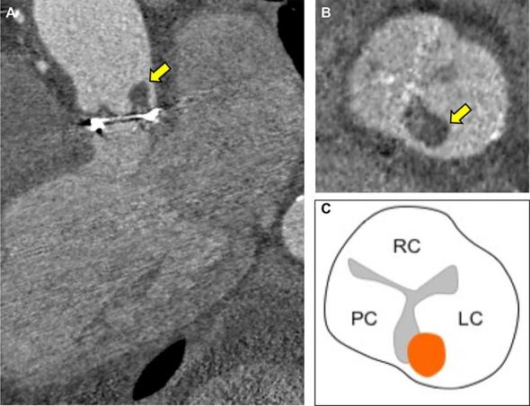
In the current era of transcatheter device therapy, the prevalence of prosthetic aortic valves and their associated complications is increasing. Echocardiography remains the first-line imaging investigation for the assessment of prosthetic valve complications, however, this often fails to identify the underlying mechanism of prosthesis failure. Recently, cardiac CT has emerged as an imaging technique capable of providing high isotropic spatial resolution of the prosthetic valve and its utility can provide important complementary diagnostic information. In this pictorial review, we present a series of common prosthetic aortic valve complications imaged with cardiac CT and demonstrate how use of this modality can enhance diagnostic accuracy.


Figures

Figure 1.
Figure 1.
TAVI hypoattenuated leaflet thickening several months after transcatheter aortic valve implantation (26 mm Spaien 3, Edwards Lifesciences). Elevated trans-prosthesis pressure gradients were observed on echocardiography and 4D cardiac CT was performed (A). Hypoattenuation of all three prosthesis cusps was present with a restricted excursion of the leaflets during ventricular systole (B). Laminar deposition of low attenuation material on the aortic aspect of the prosthesis results in the thickened appearance of the leaflet (C). Resolution with anticoagulation and restoration of normal leaflet motion is observed with this CT finding suggesting that it may represent subacute thrombus. 4D, four-dimensional; TAVI, transcatheter aortic valve implantation.
Figure 2.
Figure 2.
Bioprosthetic aortic valve pannus formation. Increased trans-prosthesis pressure gradients were recorded after surgical bioprosthetic aortic valve replacement (21 mm Carpentier-Edwards Perimount MagnaEase, Edwards Lifesciences). Sagittal multiplanar reconstruction of the aortic root (A) revealed thickening of the subvalvular apparatus propagating from the interventricular septum and aorto-mitral continuity. Axial views of the tissue at the base of the prosthesis (B) confirmed circumferential thickening below the level of the sewing ring (arrowed). The CT intensity distribution curve supported the presence of pannus (C).
Figure 3.
Figure 3.
Differentiating thrombus from pannus formation on cardiac CT. Referencing the CT attenuation of tissue to the adjacent myocardium can help differentiate low attenuation thrombus (A) from fibrotic regions of pannus (B).
Figure 4.
Figure 4.
Bioprosthetic structural valve degeneration with cusp prolapse Due to the high risk of complication from a repeat sternotomy, pre-procedural cardiac CT was performed prior to consideration for valve-in-valve TAVI in a patient with a bioprosthetic aortic valve replacement (19 mm Pericardial Elan, Vascutek). (A) Cardiac CT demonstrated failure of cusp coaptation with prolapse of the left prosthetic cusp (arrowed) into the LVOT (B), arrowed, (C), schematic representation. Cardiac CT highlighted an acquired interventricular septal aneurysm related to the angulation of the regurgitant jet. LVOT, left ventricular outflow tract; TAVI, transc atheteraortic valve implantation.
Figure 5.
Figure 5.
Bioprosthetic aortic valve endocarditis with thrombotic vegetation An elderly male presented with vertebral discitis and systemic symptoms associated with Streptococcus salivarious. 7 years previously, he had undergone surgical bioprosthetic aortic valve replacement (25 mm Mitroflow, Sorin) Transoesophageal echocardiography was unable delineate vegetations on the aortic bioprosthesis and a detailed anatomical assessment with cardiac CT was performed. (A, 3-chamber reconstruction) Cardiac CT demonstrated a low attenuation (84 HU) 8 mm lesion adhering to the commissure of the left prosthetic cusp (arrowed). (B short axis of aortic bioprosthesis with thrombotic vegetation arrowed and C, corresponding schematic representation) The low CT number is in-keeping with a thrombotic vegetation on the prosthesis. HU, Hounsfield unit.
Figure 6.
Figure 6.
Sinus of valsalva saccular pseudoaneurysm. Following a mechanical aortic valve replacement, cardiac CT was employed to measure the size and relationship of the saccular pseudoaneurysm to the great vessels and the coronaries (A). The LMCA (yellow arrow) is tented by a large saccular pseudoaneurysm that tracks along the left lateral and posterior margin of the aortic root (B) ventricular systole and (C) ventricular diastole with pseudoaneurysm. There is systolic compression (<50% luminal stenosis) of the LMCA (D, arrowed) between the common pulmonary artery and saccular aneurysm with restoration of the LMCA diameter during ventricular diastole (E, arrowed). LMCA, left main coronary artery.

References

    1. Grant SW, Hickey GL, Ludman P, Moat N, Cunningham D, de Belder M, et al. . Activity and outcomes for aortic valve implantations performed in England and Wales since the introduction of transcatheter aortic valve implantation. Eur J Cardiothorac Surg 2016; 49: 1164–73. doi: 10.1093/ejcts/ezv270 - DOI - PubMed
    1. Moss AJ, Dweck MR, Dreisbach JG, Williams MC, Mak SM, Cartlidge T, et al. . Complementary role of cardiac CT in the assessment of aortic valve replacement dysfunction. Open Heart 2016; 3: e000494. doi: 10.1136/openhrt-2016-000494 - DOI - PMC - PubMed
    1. Baumgartner H. The 2017 ESC/EACTS guidelines on the management of valvular heart disease: What is new and what has changed compared to the 2012 guidelines? Wien Klin Wochenschr 2018; 130:168–71. doi: 10.1007/s00508-017-1297-5 - DOI - PubMed
    1. Habets J, Symersky P, Leiner T, de Mol BA, Mali WP, Budde RP. Artifact reduction strategies for prosthetic heart valve CT imaging. Int J Cardiovasc Imaging 2012; 28: 2099–108. doi: 10.1007/s10554-012-0041-5 - DOI - PMC - PubMed
    1. Jilaihawi H, Asch FM, Manasse E, Ruiz CE, Jelnin V, Kashif M, et al. . Systematic CT methodology for the evaluation of subclinical leaflet thrombosis. JACC Cardiovasc Imaging 2017; 10: 461–70. doi: 10.1016/j.jcmg.2017.02.005 - DOI - PubMed