. 2018 Aug 3;14(8):e1007202.
doi: 10.1371/journal.ppat.1007202. eCollection 2018 Aug.

Inhibition of murine herpesvirus-68 replication by IFN-gamma in macrophages is counteracted by the induction of SOCS1 expression

Affiliations

Inhibition of murine herpesvirus-68 replication by IFN-gamma in macrophages is counteracted by the induction of SOCS1 expression

Yong Shen et al. PLoS Pathog. .

Abstract

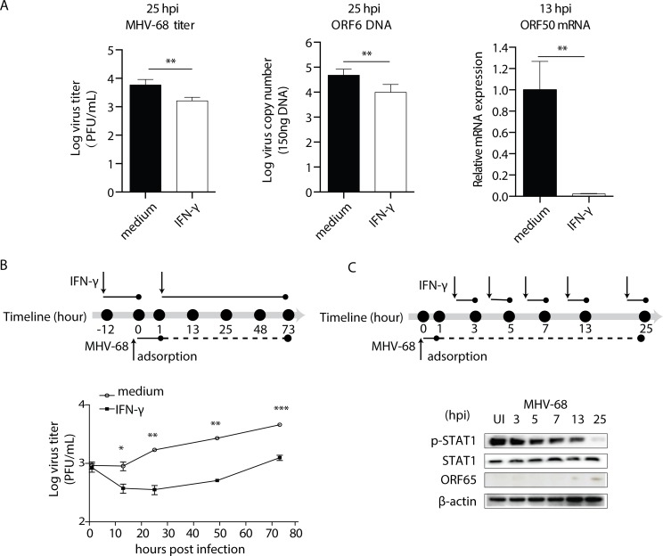
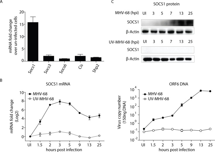
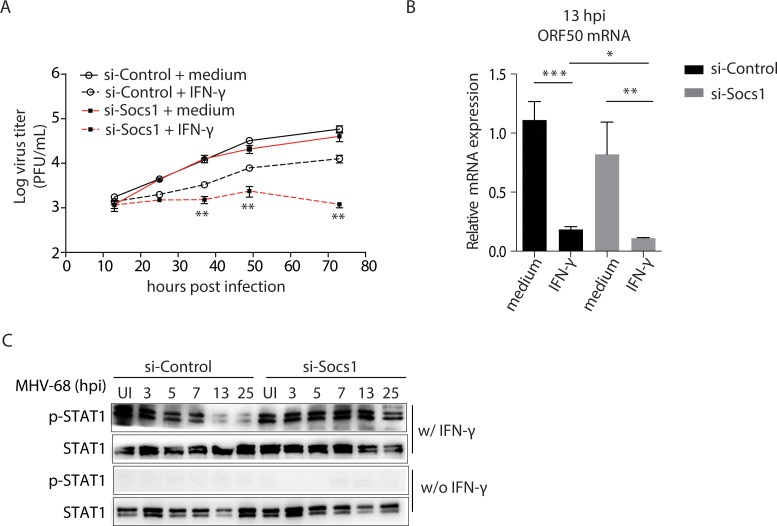
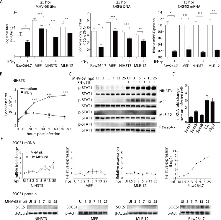
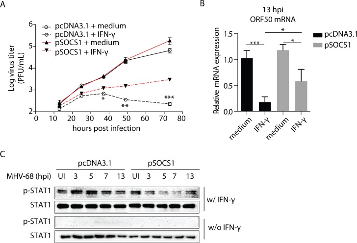
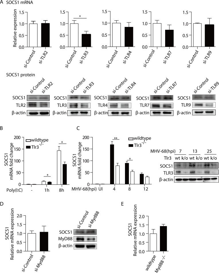
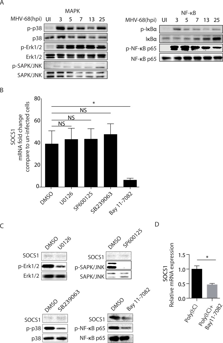
Gamma interferon (IFN-γ) is known to negatively regulate murine gammaherpesvirus-68 (MHV-68 or γHV-68) replication. This process involves the suppression of the viral gene replication and transcription activator (RTA) promoter, as well as activation of signal transducers and activators of transcription (STAT1). Notably, this effect is gradually attenuated during MHV-68 infection of bone marrow-derived macrophages (BMMs), which raised the possibility that the virus may utilize a mechanism that counteracts the antiviral effect of IFN-γ. By identifying the cellular factors that negatively regulate JAK-STAT1 signaling, we revealed that the infection of BMMs by MHV-68 induces the expression of suppressor of cytokine signaling 1 (SOCS1) and that depletion of SOCS1 restores the inhibitory effect of IFN-γ on virus replication. Moreover, we demonstrated that the expression of SOCS1 was induced as a result of the Toll-like receptor 3 (TLR3) mediated activation of the NF-κB signaling cascade. In conclusion, we report that TLR3-TRAF-NF-κB signaling pathway play a role in the induction of SOCS1 that counteracts the antiviral effect of IFN-γ during MHV-68 infection. This process is cell type-specific: it is functional in macrophages, but not in epithelial cells or fibroblasts. Our study reveals a mechanism that balances the immune responses and the escape of a gamma-herpesvirus in some antigen-presenting cells.


Conflict of interest statement

The authors have declared that no competing interests exist.

Figures

Fig 1
Fig 1. The gradual loss of the IFN-γ antiviral effect is accompanied by decreased STAT1 phosphorylation in BMMs.
(A) BMMs were pre-treated with recombinant mouse IFN-γ at 10 U/mL, or medium for 12 hours before being infected with MHV-68 at MOI = 10. At the indicated hours post infection (hpi), cells were collected for plaque assay, ORF6 DNA PCR, and ORF50 RT-qPCR to determine virus titer, viral DNA copy number, and RTA expression, respectively. The ORF50 mRNA levels are expressed as values relative to the MHV-68 infected cells without IFN-γ treatment. (B) BMMs were pre-treated with recombinant mouse IFN-γ at 10 U/mL for 12 hours before being infected with MHV-68 at MOI = 10. At the indicated hours post infection, the virus titer was determined by plaque assay. The viral growth curve was created based on the Log virus titer (PFU/mL). (C) BMMs were infected with MHV-68 at MOI = 10 and allowed to grow for the indicated periods of time. At the beginning of the last hour in each time period, 10 U/mL of IFN-γ (w/IFN-γ) was added to the culture medium. At the end of each period, the cells were collected for protein extraction, and western blotting was performed to evaluate the expression levels of STAT1 tyrosine phosphorylation (p-STAT1) and STAT1. At the same times, ORF65 western blotting was performed to measure MHV-68 protein expression levels. The results are representative of three independent experiments and are expressed as means ± S.E.M. *p < 0.05, **p < 0.01, ***p < 0.001.
Fig 2
Fig 2. MHV-68 induces SOCS1 expression in BMMs.
(A) BMMs were infected with MHV-68 at MOI = 10. At 13 hpi, total RNA was extracted and the mRNAs levels of Socs1, Socs3, Socs6, Cis and Shp2 were determined by RT-qPCR. (B-C) BMMs were either infected with MHV-68 or UV-inactivated MHV-68. At the indicated hours post infection, the cells were collected for RT-qPCR and western blotting to evaluate the SOCS1 mRNA and protein expression levels. In addition, ORF6 DNA qPCR was performed to assess the extent of viral replication. SOCS1 qPCR data are expressed as fold change in mRNA level compared to that in uninfected cells. The results are representative of three independent experiments and are expressed as means ± S.E.M.
Fig 3
Fig 3. Knock down of SOCS1 results in a persistent antiviral effect of IFN-γ and tyrosine phosphorylation of STAT1 during MHV-68 infection.
BMM cells were transfected with an siRNA against SOCS1 (si-Socs1) or an irrelevant target (si-Control). (A) At 24 hours after transfection, the cells were pre-treated with IFN-γ (10 U/mL) for 12 hours, and were then infected with MHV-68 at MOI = 10. At the indicated times post infection, the viral titer was measured by plaque assay. **p < 0.01, si-Socs1+IFN-γ vs. si-Control+IFN-γ. (B) At 13 hpi, viral ORF50 mRNA expression was detected by RT-qPCR. Data are expressed as relative values to the MHV-68 infected, si-Control transfected cells without IFN-γ treatment. *p < 0.05, **p < 0.01, ***p < 0.001. (c) Cells were infected with MHV-68 at MOI = 10 and allowed to grow for the indicated time periods. At the beginning of the last hour in each time period, 10 U/mL of IFN-γ (w/IFN-γ) or medium (w/o IFN-γ) was added to the culture medium. At the end of each time period, western blotting was performed to evaluate the expression levels of STAT1 tyrosine phosphorylation (p-STAT1) and STAT1. Results are representative of three independent experiments and are expressed as mean ± S.E.M.
Fig 4
Fig 4. In NIH3T3 fibroblasts, MHV-68 infection does not lead to SOCS1 expression.
(A) IFN-γ inhibits the lytic replication of MHV-68 in Raw264.7, MEF, NIH3T3, and MLE-12 cells. Raw264.7, MEF, NIH3T3, and MLE-12 cells were pre-treated with IFN-γ at 10 U/mL for 12 hours, and were then infected with MHV-68 at MOI = 10 (Raw264.7) or 1 (MEF, NIH3T3, MLE-12). At the indicated hours post infection (hpi), the cells were collected for plaque assay, ORF6 DNA PCR, and ORF50 RT-qPCR to evaluate virus titer, viral DNA copy number, and RTA expression, respectively. The ORF50 mRNA levels are expressed as values relative to the MHV-68 infected cells without IFN-γ treatment. (B) The antiviral role of IFN-γ is stable in NIH3T3 cells. NIH3T3 cells were pre-treated with IFN-γ at 10 U/mL for 12 hours before being infected with MHV-68 at MOI = 10. At the indicated hours post infection, the virus titer was determined by plaque assay. The viral growth curve was created based on Log virus titer (PFU/mL). (C) IFN-γ induced tyrosine phosphorylation of STAT1 is stable in NIH3T3, MEF, and MLE-12 cells, but gradually declines in Raw264.7 cells. NIH3T3, MEF, MLE-12, and Raw264.7 cells were infected with MHV-68 and allowed to grow for the indicated periods of time. At the beginning of last hour in each time period, 10 U/mL of IFN-γ was added to the culture medium. At the end of each time period, cells were collected for protein extraction, and western blotting was performed to evaluate the expression levels of p-STAT1 and STAT1. (D) MHV-68 does not induce Socs1, Socs3, Socs6, Cis, or Shp2 mRNA expression in NIH3T3 cells. NIH3T3 cells were infected with MHV-68 at MOI = 1. At 13 hpi, total RNA was extracted and the mRNAs of Socs1, Socs3, Socs6, Cis, and Shp2 were detected by RT-qPCR. (E) MHV-68 does not induce SOCS1 mRNA or protein expression in NIH3T3, MEF, and MLE-12 cells but induces both SOCS1 mRNA and protein expressions in Raw264.7 cells. Cells were either infected with MHV-68 or UV-inactivated MHV-68. At the indicated hours post infection, cells were collected for RT-qPCR and western blotting to evaluate SOCS1 mRNA and protein expression. qPCR data are expressed as fold change in mRNA level compared to that in uninfected cells. Results are representative of three independent experiments and are expressed as mean ± S.E.M., *p < 0.05, **p < 0.01, ***p < 0.001.
Fig 5
Fig 5. Overexpression of SOCS1 decreases the antiviral effect of IFN-γ and tyrosine phosphorylation of STAT1 during MHV-68 infection in NIH3T3 fibroblasts.
NIH3T3 cells were transfected with an expression plasmid encoding SOCS1 (pSOCS1) or control (pcDNA3.1). (A) At 48 hours after transfection, the cells were pre-treated with IFN-γ (10 U/mL) for 12 hours, and were then infected with MHV-68 at MOI = 10. At the indicated times post infection, the viral titer was measured by plaque assay. *p < 0.05, **p < 0.01, ***p<0.001, pSOCS1+IFN-γ vs. pcDNA3.1+IFN-γ. In parallel, western blotting was performed to assess the SOCS1 expression levels in cells without IFN-γ treatment. (B) RT-qPCR was used to detect the viral ORF50 mRNA expression at 13 hpi. Data are expressed as values relative to the MHV-68 infected, pcDNA3.1 transfected cells without IFN-γ treatment. *p < 0.05, ***p < 0.001. (C) Cells were infected with MHV-68 for the indicated times, with the last hour of infection being in the presence of IFN-γ (10 U/mL) or not. Proteins were extracted, and western blotting was performed to evaluate the tyrosine phosphorylation level of STAT1. Results are representative of three independent experiments and are expressed as mean ± S.E.M.
Fig 6
Fig 6. TLR3 mediates the MHV-68-induced SOCS1 production.
(A) BMMs were transfected with siRNAs against Tlr2, Tlr3, Tlr4, Tlr7, Tlr9 (si-TLR) or an irrelevant target (si-Control), respectively. Forty-eight hours post transfection, cells were infected with MHV-68 at MOI = 10. At 8 hpi, RNA and protein were then extracted. RT-qPCR was performed to determine SOCS1 mRNA levels and western blotting was performed to determine the protein expression levels of SOCS1 and the individual TLRs, respectively. The SOCS1 mRNA levels are expressed as values relative to the MHV-68 infected, si-Control transfected cells. (B) BMMs from Tlr3-/- and wildtype C57BL/6 mice were treated with poly(I:C) at 10 μg/mL for 1 and 8 hours, respectively. Total RNA was extracted and SOCS1 mRNA levels were determined by RT-qPCR. qPCR data are expressed as fold change in mRNA level compared to that in untreated WT cells. (C) BMMs from Tlr3-/- mice and wild type (WT) mice were infected MHV-68. At indicated hpi, cells were harvested to measure the SOCS1 mRNA levels by the RT-qPCR, and to measure the TLR3 and SOCS1 proteins levels by western blotting. qPCR data were expressed as fold change in mRNA level compared to that in uninfected WT cells. (D) BMMs were transfected with si-MyD88 or si-Control. Forty-eight hours post-transfection, the cells were infected with MHV-68. At 8 hpi, the SOCS1 mRNA levels and protein levels were determined. The SOCS1 mRNA levels are expressed as values relative to the MHV-68 infected, si-Control transfected cells. (E) BMMs from Myd88-/- mice and wildtype C57BL/6 mice were infected with MHV-68. At 8 hpi, total RNA was extracted and the mRNA expression of SOCS1 was determined by RT-qPCR. The SOCS1 mRNA levels are expressed as values relative to the MHV-68 infected WT cells. Results are representative of three independent experiments and are expressed as mean ± S.E.M., *p < 0.05.
Fig 7
Fig 7. NF-κB is responsible for the MHV-68-induced SOCS1 production.
(A) BMMs were infected with MHV-68 at MOI = 10. At 3, 5, 7, 13, and 25 hpi, protein was extracted and the levels of phospho-p38, p38, phospho-Erk1/2, Erk1/2, phospho-SAPK/JNK, SAPK/JNK, phospho-IκBα, IκBα, phospho-NF-κB p65, NF-κB p65 were determined by western blotting. (B) BMMs were pretreated with DMSO, U0126, SP600125, SB239063 or Bay11-7082, respectively for 30 min before being infected with MHV-68. At 8 hpi, SOCS1 mRNA was determined by RT-qPCR. Data are expressed as fold change in mRNA level compared to that in uninfected cells. (C) BMMs were pretreated with U0126, SP600125, SB239063, or Bay11-7082 for 30 min before being infected with MHV-68. At 8 hpi, the levels of SOCS1, phospho-NF-κB p65, NF-κB p65, phospho-Erk1/2, Erk1/2, phospho-p38, p38, phospho-SAPK/JNK, and SAPK/JNK were determined by western blotting. (D) BMM cells were pretreated with Bay11-7082 for 30 min before cells were stimulated with Poly(I:C) for 12 hours. Cells were collected for RNA extraction and SOCS1 expression was determined by RT-qPCR. Data are expressed as SOCS1 relative mRNA values over Poly(I:C) stimulated cells without inhibitor treatment. Results are representative of three independent experiments and are expressed as mean ± S.E.M., *p < 0.05.

Similar articles

Cited by

References

    1. Efstathiou S, Ho YM, Hall S, Styles CJ, Scott SD, et al. (1990) Murine herpesvirus 68 is genetically related to the gammaherpesviruses Epstein-Barr virus and herpesvirus saimiri. J Gen Virol 71 (Pt 6): 1365–1372. - PubMed
    1. Virgin HWt, Latreille P, Wamsley P, Hallsworth K, Weck KE, et al. (1997) Complete sequence and genomic analysis of murine gammaherpesvirus 68. J Virol 71: 5894–5904. - PMC - PubMed
    1. Martinez-Guzman D, Rickabaugh T, Wu TT, Brown H, Cole S, et al. (2003) Transcription program of murine gammaherpesvirus 68. J Virol 77: 10488–10503. 10.1128/JVI.77.19.10488-10503.2003 - DOI - PMC - PubMed
    1. Stewart JP, Usherwood EJ, Ross A, Dyson H, Nash T (1998) Lung epithelial cells are a major site of murine gammaherpesvirus persistence. J Exp Med 187: 1941–1951. - PMC - PubMed
    1. Leang RS, Wu TT, Hwang S, Liang LT, Tong L, et al. (2011) The anti-interferon activity of conserved viral dUTPase ORF54 is essential for an effective MHV-68 infection. PLoS Pathog 7: e1002292 10.1371/journal.ppat.1002292 - DOI - PMC - PubMed

Publication types

MeSH terms

Substances