. 2018 Aug 3;6(1):72.
doi: 10.1186/s40478-018-0579-0.

Novel antibodies reveal presynaptic localization of C9orf72 protein and reduced protein levels in C9orf72 mutation carriers

Affiliations

Novel antibodies reveal presynaptic localization of C9orf72 protein and reduced protein levels in C9orf72 mutation carriers

Petra Frick et al. Acta Neuropathol Commun. .

Abstract

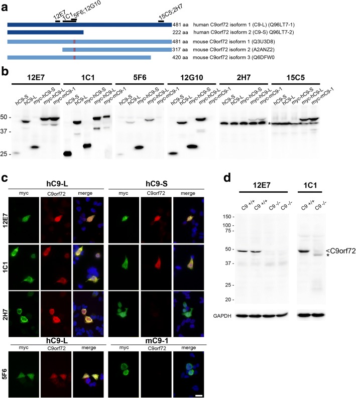
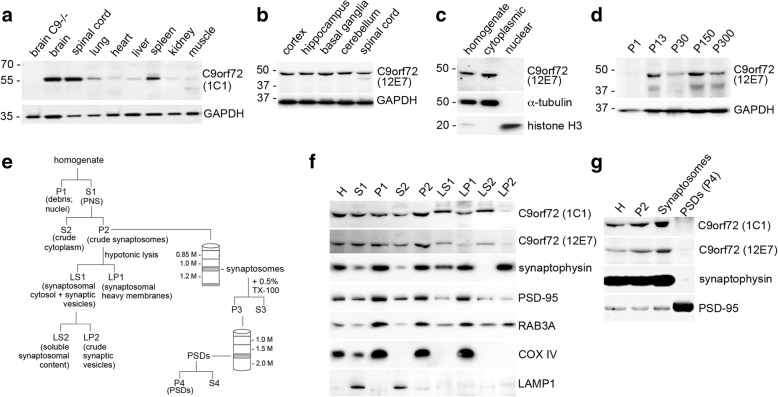
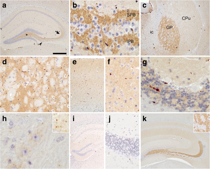
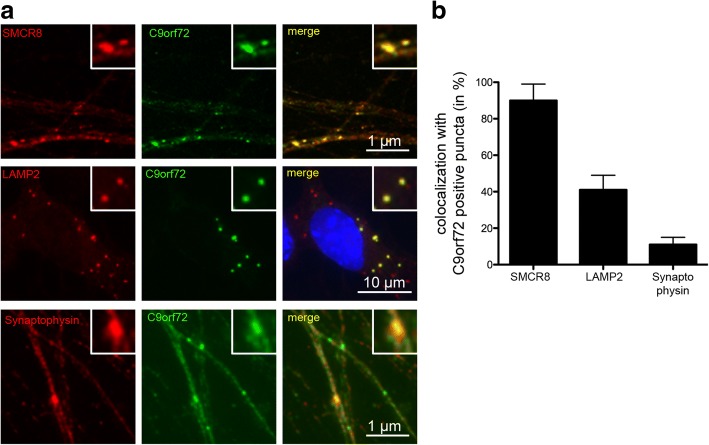
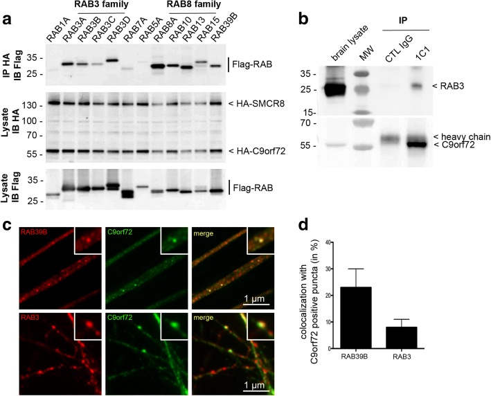
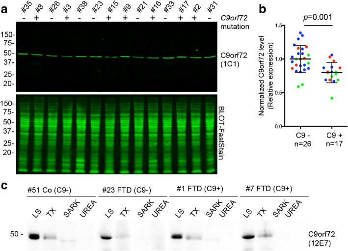
Hexanucleotide repeat expansion in C9orf72 is the most common genetic cause of frontotemporal dementia and amyotrophic lateral sclerosis, but the pathogenic mechanism of this mutation remains unresolved. Haploinsufficiency has been proposed as one potential mechanism. However, insights if and how reduced C9orf72 proteins levels might contribute to disease pathogenesis are still limited because C9orf72 expression, localization and functions in the central nervous system (CNS) are uncertain, in part due to the poor specificity of currently available C9orf72 antibodies.Here, we generated and characterized novel knock-out validated monoclonal rat and mouse antibodies against C9orf72. We found that C9orf72 is a low abundant, cytoplasmic, highly soluble protein with the long 481 amino acid isoform being the predominant, if not exclusively, expressed protein isoform in mouse tissues and human brain. As consequence of the C9orf72 repeat expansion, C9orf72 protein levels in the cerebellum were reduced to 80% in our series of C9orf72 mutation carriers (n = 17) compared to controls (n = 26). However, no associations between cerebellar protein levels and clinical phenotypes were seen. Finally, by utilizing complementary immunohistochemical and biochemical approaches including analysis of human iPSC derived motor neurons, we identified C9orf72, in addition to its association to lysosomes, to be localized to the presynapses and able to interact with all members of the RAB3 protein family, suggestive of a role for C9orf72 in regulating synaptic vesicle functions by potentially acting as guanine nucleotide exchange factor for RAB3 proteins.In conclusion, our findings provide further evidence for haploinsufficiency as potential mechanism in C9orf72 pathogenesis by demonstrating reduced protein levels in C9orf72 mutation carriers and important novel insights into the physiological role of C9orf72 in the CNS. Moreover, the described novel monoclonal C9orf72 antibodies will be useful tools to further dissect the cellular and molecular functions of C9orf72.

Keywords: Amyotrophic lateral sclerosis; C9orf72; Frontotemporal dementia; Frontotemporal lobar degeneration; RAB3; Synaptic vesicles.


Conflict of interest statement

The authors declare that they have no competing interests

Springer Nature remains neutral with regard to jurisdictional claims in published maps and institutional affiliations.

Figures

Fig. 1
Fig. 1
Basic characterization of novel monoclonal antibodies against C9orf72. a Schematic representation of postulated human and murine C9orf72 protein isoforms with epitopes recognized by novel monoclonal antibodies (mAbs) against C9orf72. In humans, two C9orf72 protein isoforms are postulated with isoform 1 representing a 481 amino acid protein, also known as long isoform or C9-L (transcribed by transcript variant 2 with the GGGGCC repeat located in the promoter region and transcript variant 3 with the GGGGCC repeat located in the first intron); and isoform 2 representing a 222 amino acid protein, also known as short isoform or C9-S (transcribed by transcript variant 1 with the GGGGCC repeat located in the first intron). In mice, three protein isoforms are postulated, with isoform 1 corresponding in size to human C9-L with 98% similarity on amino acid sequence. The red lines in the murine isoforms illustrate two amino acid changes between the human and mouse C9orf72 sequence in the epitope recognized by mAbs 5F6 and 12G10. b Immunoblot analysis of protein lysates of HEK293 cells expressing untagged or myc-DDK-tagged human C9-L and C9-S or myc-DDK-tagged murine C9orf72 isoform 1 (mC9–1) with novel C9orf72 mAbs. Clones 12E7 and 1C1 recognize hC9-S and hC9-L as well as mC9–1. Clones 5F6 and 12G10 specifically recognize human but not mouse C9orf72. Clones 2H7 and 15C5 specifically recognize an epitope in the C-terminus only present in hC9-L and mC9–1 but not hC9-S, however, both mAbs also recognize an unspecific band (asterisk). c Double label immunofluorescence for anti-myc (green) and anti-C9orf72 (red) of HEK293 cells transiently expressing myc-DDK-tagged hC9-L, hC9-S or mC9–1 confirms the specificity of the indicated mAbs for specific C9orf72 isoforms or species. Hoechst 33342 staining of nuclei (blue) in the merged images. Scale bar: 20 μm. d Immunoblot analysis of total protein lysates from brains of wild-type (C9+/+) and C9orf72 knock-out (C9−/−) mice. Only a single band around 50 kDa corresponding in size to the murine isoform 1 is detected with mAbs 12E7 and 1C1 in wild-type mice (arrowhead). Note, that this band is completely absent in C9−/− mice, validating the high specificity for C9orf72 of the mAbs 12E7 and 1C1. The weak band labeled with an asterisk seen in C9−/− with the mouse mAb 1C1 represents mouse IgG heavy chain recognized by the anti-mouse IgG (H + L) detection antibody (see Additional file 1: Figure S1b for secondary antibody control). GAPDH is shown as loading control. MW size marker: Precision Plus Protein Dual Color Standards (b and d)
Fig. 2
Fig. 2
C9orf72 is enriched in synaptosomes. a Immunoblot analysis of total protein lysates of different mouse tissues reveals widespread C9orf72 protein expression detected as single band around 50 kDa with highest expression levels in brain followed by spinal cord. GAPDH is shown as loading control. b No obvious changes are observed between different brain regions by immunoblot analysis of protein lysates from cortex, hippocampus, striatum, cerebellum and spinal cord. GAPDH is shown as loading control. c Immunoblot analysis of nuclear and cytoplasmic protein fractionations extracted from adult mouse brain reveals localization of C9orf72 to the cytoplasm. α-tubulin and Histone H3 are shown to demonstrate purity of the cytoplasmic and nuclear fractions, respectively. d Immunoblot analysis and quantification of C9orf72 expression levels over mouse brain development from P1 to P300 showing increase of C9orf72 between P1 and P16. e Schematic of the purification protocols for synaptic vesicles and postsynaptic densities (PSDs). Mouse forebrains were homogenized and centrifuged to generate a crude synaptosomal fraction (P2). P2 fractions were fractionated into synaptosomal heavy membranes (LP1), synaptic vesicles (LP2), and synaptic cytosol (LS2) by hypotonic lysis and differential centrifugation. Alternatively, P2 fractions were centrifuged through sucrose gradient to reveal a pure synaptosomal fraction which was further processed to isolate pure PSDs. f C9orf72 is detectable in the synaptosomal fraction P2, and is released into the LS2 fraction containing the soluble cytoplasmic content of synaptosomes. g C9orf72 is enriched in the pure synaptosomal fraction, but absent from PSD fractions. f and g The purity of fractions was confirmed with specific marker proteins for synaptic vesicles (synaptophysin), postsynaptic densities (PSD-95), mitochondria (Cox-IV), and lysosomes (Lamp1). MW size marker: PageRule Plus Prestained Protein Ladder (a); Precision Plus Protein Dual Color Standards (b-d, f, g)
Fig. 3
Fig. 3
C9orf72 immunohistochemistry reveals synaptic staining pattern with enrichment in hippocampal mossy fiber terminals. Immunohistochemistry with anti-C9orf72 mAb 1C1 (a-j); immunohistochemistry with anti-synaptoporin antibody (k). In the adult mouse brain (a-h), strong immunoreactivity for C9orf72 is seen in the hippocampal mossy fiber system (a) with labeling in the hilus (asterisk), stratum lucidum (arrow) and infrapyramidal mossy fiber bundles (arrowhead). (b) Higher magnification of punctate staining pattern of mossy fiber terminals in suprapyramidal (SPB) and infrapyramidal (IPB) mossy fiber bundles. Robust staining was also observed in the globus pallidus (GP) (c and d) while the caudate putamen (CPu) (c) and other gray matter regions showed weaker immunoreactivity of the neuropil as shown for frontal cortex (e and f) and cerebellum with predominant staining in the molecular and granular layer (g). No immunoreactivity is seen in the white matter and internal capsule (ic). In addition to punctate neuropil staining, neurons with large cytoplasm such as motor neurons in the spinal cord showed several cytoplasmic puncta (h). (i and j): Specificity of anti-C9orf72 immunohistochemistry was validated by the complete absence of immunoreactivity in brain sections from C9orf72 knock-out mice as shown for hippocampus (i) and cerebellum (j). Note the strikingly similar staining pattern of the mossy fiber terminals in the hippocampus for C9orf72 (a) and for the presynaptic marker protein synaptoporin (k). Scale bar: 533 μm (c); 400 μm (a, i, k); 267 μm (e); 80 μm (d, j, insert k); 40 μm (b, f, g); 20 μm (h); 6,5 μm (insert h)
Fig. 4
Fig. 4
C9orf72 co-localizes with synaptic vesicles in human iPSC derived motor neurons. a C9orf72 positive puncta (green) are seen in the axons of 30 day old human iPSC derived motor neurons which consistently co-localize with SMCR8 (red, upper panel) and partially co-localize with LAMP2 as lysosomal marker (red, middle panel) or with the synaptic vesicle marker synaptophysin (red, lower panel). Nuclei are stained with DAPI (blue) in the merged images. C9orf72 labeled with 12E7 antibody in the upper panel and with 1C1 in the middle and lower panel. b Graph showing the percentage of C9orf72 positive puncta co-localizing with SMCR8, LAMP2 or synaptophysin. Values are shown as mean ± SD
Fig. 5
Fig. 5
C9orf72 complex interacts with members of the RAB3 protein family. a Immunoblot analysis of HA-immunoprecipitated proteins from lysates of HEK293 cells co-expressing HA-tagged human C9orf72 and HA-tagged SMCR8 with various FLAG-tagged Rabs showing co-immunoprecipitation of all members of the RAB3 family with the C9orf72 complex. Other Rabs were used as positive (RAB8 subfamily, RAB39B) or negative (RAB1A, RAB7A, RAB5A) controls based on published reports. b Immunoblot against RAB3 of control (IgG alone) or endogenous C9orf72 immunoprecipitated proteins from lysates of adult mouse brain. MW size marker: PageRule Plus Prestained Protein Ladder (a and b). c Double-label immunofluorescence of 30 day old human iPSC-derived motor neurons showing co-localization of C9orf72 (green) with RAB39B (red, upper panel) or RAB3 (red, lower panel) in a subset of C9orf72-positive puncta. C9orf72 labeled with 12E7 in the upper panel and 1C1 in the lower panel. d Graph showing the percentage of C9orf72 positive puncta co-localizing with RAB3 or RAB39B. Values are shown as mean ± SD
Fig. 6
Fig. 6
Reduced C9orf72 expression levels in the cerebellum of C9orf72 mutation carriers. a Immunoblot analysis of C9orf72 protein levels in RIPA lysates extracted from frozen cerebellar gray matter of C9orf72 mutation cases and neurologic controls reveals a single band ~ 50 kDa corresponding in size to the long 481 amino acid isoform of C9orf72 (C9-L). Total protein stains are shown as loading controls. The blot shown is representative of three independent experiments. b Quantification of C9orf72 protein levels in the cerebellum of n = 17 cases with C9orf72 repeat expansions (C9+) and n = 26 controls (C9-). Dot blot of normalized C9orf72 values with mean and standard deviation shown as line and error bars. Different colors represent clinical phenotypes (green = FTD; red = ALS/FTD; blue = ALS). p = 0.001 by Student’s two-tailed, unpaired t test. c Proteins were sequentially extracted from frozen frontal cortex of C9orf72 mutation carriers (C9+) and controls (C9-) with a series of buffers of increasing stringency to receive low salt (LS), high-salt Triton-X-100 (TX), sarkosyl (SARK), and urea protein fractions for immunoblot analysis. Human C9orf72 (C9-L) is present in all cases in the fractions enriched for highly soluble proteins (LS and to lesser extent TX) with no changes observed in solubility between C9+ and C9- cases. MW size marker: PageRule Plus Prestained Protein Ladder (a and c)

References

    1. Amick J, Roczniak-Ferguson A, Ferguson SM. C9orf72 binds SMCR8, localizes to lysosomes, and regulates mTORC1 signaling. Mol Biol Cell. 2016;27:3040–3051. doi: 10.1091/mbc.e16-01-0003. - DOI - PMC - PubMed
    1. Ash PE, Bieniek KF, Gendron TF, Caulfield T, Lin WL, Dejesus-Hernandez M, et al. Unconventional translation of C9ORF72 GGGGCC expansion generates insoluble polypeptides specific to c9FTD/ALS. Neuron. 2013;77:639–646. doi: 10.1016/j.neuron.2013.02.004. - DOI - PMC - PubMed
    1. Atanasio A, Decman V, White D, Ramos M, Ikiz B, Lee HC, et al. C9orf72 ablation causes immune dysregulation characterized by leukocyte expansion, autoantibody production, and glomerulonephropathy in mice. Sci Rep. 2016;6:23204. doi: 10.1038/srep23204. - DOI - PMC - PubMed
    1. Atkinson RA, Fernandez-Martos CM, Atkin JD, Vickers JC, King AE. C9ORF72 expression and cellular localization over mouse development. Acta Neuropathol Commun. 2015;3:59. doi: 10.1186/s40478-015-0238-7. - DOI - PMC - PubMed
    1. Bertrand A, Wen J, Rinaldi D, Houot M, Sayah S, Camuzat A et al (2017) Early cognitive, structural, and microstructural changes in Presymptomatic C9orf72 carriers younger than 40 years. JAMA Neurol. 10.1001/jamaneurol.2017.4266 - PMC - PubMed

Publication types

MeSH terms