. 2018 Aug 3;11(1):558.
doi: 10.1186/s13104-018-3664-3.

Genetic and genomic stability across lymphoblastoid cell line expansions

Affiliations

Genetic and genomic stability across lymphoblastoid cell line expansions

Laura B Scheinfeldt et al. BMC Res Notes. .

Abstract

Objective: Lymphoblastoid cell lines are widely used in genetic and genomic studies. Previous work has characterized variant stability in transformed culture and across culture passages. Our objective was to extend this work to evaluate single nucleotide polymorphism and structural variation across cell line expansions, which are commonly used in biorepository distribution. Our study used DNA and cell lines sampled from six research participants. We assayed genome-wide genetic variants and inferred structural variants for DNA extracted from blood, from transformed cell cultures, and from three generations of expansions.

Results: Single nucleotide variation was stable between DNA and expanded cell lines (ranging from 99.90 to 99.98% concordance). Structural variation was less consistent across expansions (median 33% concordance) with a noticeable decrease in later expansions. In summary, we demonstrate consistency between SNPs assayed from whole blood DNA and LCL DNA; however, more caution should be taken in using LCL DNA to study structural variation.

Keywords: Copy number variant; Genomic; Lymphoblastoid cell line; Single nucleotide polymorphism; Stability; Structural variation.


Figures

Fig. 1
Fig. 1
Diagram of cell transformation, passaging and expansion. Whole blood is collected from research participants, and peripheral blood mononuclear cells are isolated and transformed into lymphoblastoid cell lines (LCLs) with Epstein Barr Virus. LCLs are grown in medium as growth plateaus due to high cell density and prior to over-saturation. Passaging (splitting the culture into diluted sub-cultures) enables LCLs to continue to grow over time. To reach expansion volumes, sub-cultured cells are combined into a larger container and diluted with medium to enable further growth (second passage). When the culture again reaches high cell density at high volume the expanded cells are harvested for cryopreservation and made available for DNA or LCL distribution
Fig. 2
Fig. 2
SNP concordance across cell culture expansions. This figure displays boxplots of the distribution of pairwise concordance between DNA (from blood) and cell culture for each individual subject. Concordance for the initial transformed cell culture and three expansions cultures are shown along the X-axis. The Y-axis displays the percentage of SNPs that were estimated to be shared using the identity by state method [13]. Red dash lines indicate the minimum and maximum SNP concordance across technical replicates
Fig. 3
Fig. 3
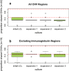
CNV concordance across cell culture expansions. This figure displays boxplots of the distribution of pairwise shared CNV regions between DNA (from blood) and cell culture for each individual subject. Shared CNV regions for the initial transformed cell culture and three expansions cultures are shown along the X-axis. The Y-axis displays the percentage of CNV regions that were estimated to be shared using PennCNV calls [11]. Red dash lines indicate the minimum and maximum number of shared CNVs across technical replicates. a Displays results for all CNV regions, and b displays results that exclude the three immunoglobulin regions identified in the samples

References

    1. Caliskan M, Pritchard JK, Ober C, Gilad Y. The effect of freeze-thaw cycles on gene expression levels in lymphoblastoid cell lines. PLoS ONE. 2014;9(9):e107166. doi: 10.1371/journal.pone.0107166. - DOI - PMC - PubMed
    1. Shim SM, Nam HY, Lee JE, Kim JW, Han BG, Jeon JP. MicroRNAs in human lymphoblastoid cell lines. Crit Rev Eukaryot Gene Expr. 2012;22(3):189–196. doi: 10.1615/CritRevEukarGeneExpr.v22.i3.20. - DOI - PubMed
    1. Moen EL, Godley LA, Zhang W, Dolan ME. Pharmacogenomics of chemotherapeutic susceptibility and toxicity. Genome Med. 2012;4(11):90. doi: 10.1186/gm391. - DOI - PMC - PubMed
    1. Hussain T, Mulherkar R. Lymphoblastoid cell lines: a continuous in vitro source of cells to study carcinogen sensitivity and DNA repair. Int J Mol Cell Med. 2012;1(2):75–87. - PMC - PubMed
    1. Barrett R, Ornelas L, Yeager N, Mandefro B, Sahabian A, Lenaeus L, Targan SR, Svendsen CN, Sareen D. Reliable generation of induced pluripotent stem cells from human lymphoblastoid cell lines. Stem Cells Transl Med. 2014;3(12):1429–1434. doi: 10.5966/sctm.2014-0121. - DOI - PMC - PubMed