. 2018 Aug;20(8):1089-1101.
doi: 10.1016/j.jcyt.2018.05.007. Epub 2018 Jul 31.

Rapid ex vivo expansion of highly enriched human invariant natural killer T cells via single antigenic stimulation for cell therapy to prevent graft-versus-host disease

Affiliations

Rapid ex vivo expansion of highly enriched human invariant natural killer T cells via single antigenic stimulation for cell therapy to prevent graft-versus-host disease

Abel Trujillo-Ocampo et al. Cytotherapy. 2018 Aug.

Abstract

Background aims: CD1d-restricted invariant natural killer (iNK) T cells are rare regulatory T cells that may contribute to the immune-regulation in allogeneic stem cell transplantation (ASCT). Here, we sought to develop an effective strategy to expand human iNK T cells for use in cell therapy to prevent graft-versus-host disease (GVHD) in ASCT.

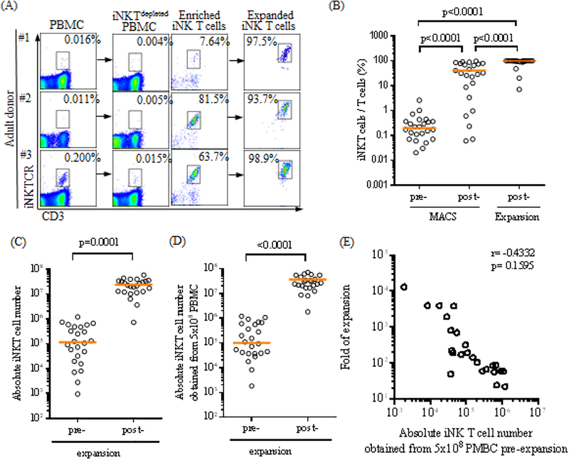
Methods: Human iNK T cells were first enriched from peripheral blood mononuclear cells (PBMCs) using magnetic-activated cell sorting separation, then co-cultured with dendritic cells in the presence of agonist glycolipids, alpha-galactosylceramide, for 2 weeks.

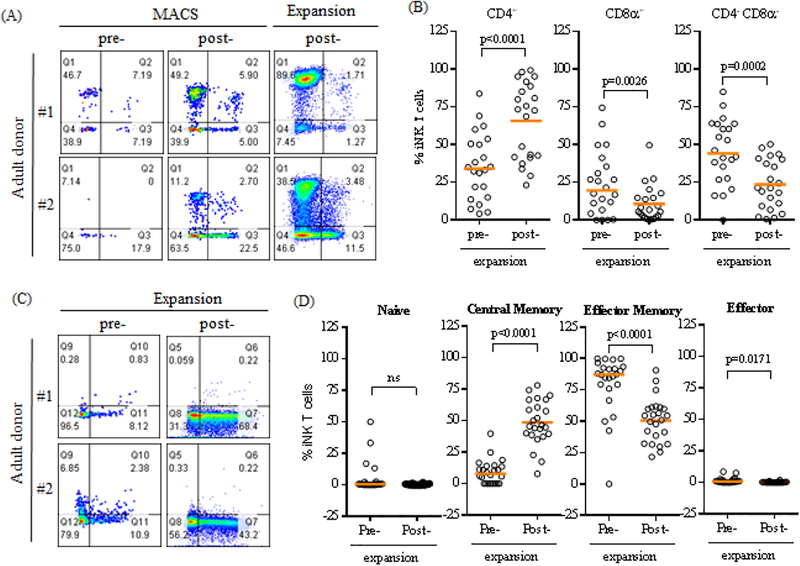
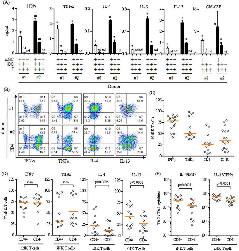
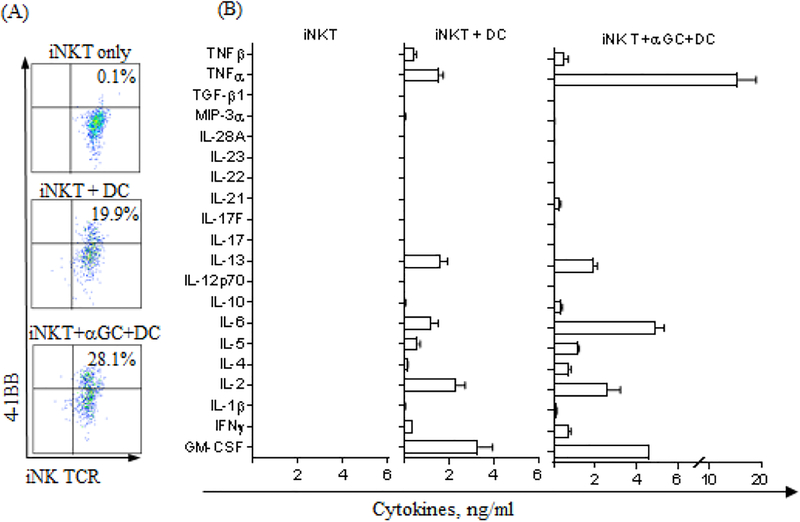
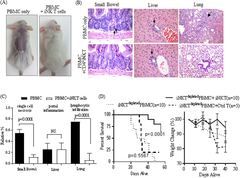
Results: The single antigenic stimulation reliably expanded iNK T cells to an average of 2.8 × 10⁷ per 5 × 10⁸ PBMCs in an average purity of 98.8% in 2 weeks (N = 24). The expanded iNK T cells contained a significantly higher level of CD4+ and central memory phenotype (CD45RA-CD62L+) compared with freshly isolated iNK T cells, and maintained their ability to produce both Th-1 (interferon [IFN]γ and tumor necrosis factor [TNF]α) and Th-2 type cytokines (interleukin [IL]-4, IL-5 and IL-13) upon antigenic stimulation or stimulation with Phorbol 12-myristate 13-acetate/ionomycin. Interestingly, expanded iNK T cells were highly autoreactive and produced a Th-2 polarized cytokine production profile after being co-cultured with dendritic cells alone without exogenous agonist glycolipid antigen. Lastly, expanded iNK T cells suppressed conventional T-cell proliferation and ameliorated xenograft GVHD (hazard ratio, 0.1266; P < 0.0001).

Conclusion: We have demonstrated a feasible approach for obtaining ex vivo expanded, highly enriched human iNK T cells for use in adoptive cell therapy to prevent GVHD in ASCT.

Keywords: cell therapy; ex vivo expansion; graft-versus-host disease; human iNK T cells.


Figures

Figure 1.
Figure 1.
A single antigenic stimulation efficiently expands highly pure human iNK T cells. (A-B) The iNK T cells were enriched from adult peripheral blood mononuclear cells (PBMC) via anti-iNKT microbeads/MACS separation, and subsequently co-cultured with allogeneic dendritic cells (DC) in the presence of αGalCer and IL-2 for 10–14 days. The representative flow cytometric analyses of iNK T cells pre and post MACS separation and post expansion from three donors were shown in (A), and percentages of iNK T cells from total T cells at each step from 24 consecutive donors were shown in (B). The purity of iNK T cells increased from average (median, hereafter) 0.195% pre-MACS separation to an average of 31.9% post MACS separation, and to 98.8% after a single round of antigen specific expansion (n=24). Absolute numbers of iNK T cells obtained before and after the expansion from 24 consecutive donors are shown in (C), and these values were adjusted to 5×10⁸ PBMC (D). Folds of expansion were plotted against absolute numbers of iNK T cells obtained from 5×10⁸ PMBC prior to the expansion (E). A total of 1.1×10⁵ iNK T cells on average (range: 948 to 1.2×10⁶) were obtained from 2×10⁸ to 1×10⁹ PBMC from 24 consecutive donors, and underwent an average of 156 folds of expansion (range 22 to 13186), resulting in an average of 2.1×10⁷ iNK T cells (range: 7.2×10⁵ to 5.7×10⁷). When these values were adjusted to 5×10⁸ PBMC, the average of 1.0×10⁵ iNK T cells (range: 1823 to 1.2×10⁶) were obtained from 5×10⁸ PBMC, and an average of 2.8×10⁷ iNK T cells (range: 1.8×10⁶ to 1.7×10⁸) were obtained after the expansion. There was a trend towards achieving the higher folds of expansion when the lower absolute number of iNK T cells were obtained prior to the expansion, but this was not statistically significant. A single symbol represents a value from a single donor. The median was used as the average value. A paired student t-test was used to compare the differences between selected groups. P-value less than 0.05 was considered as “significant”
Figure 2.
Figure 2.
A single antigen stimulation preferentially expand CD4+ and CD45RACD62L+ iNK T cells. (A) The representative flow cytometric analysis of CD4 and CD8α expression on iNK T cells pre- and post- enrichment, and post- expansion. (B) The percent CD4+, CD8α+, CD4CD8α iNK T cells pre- and post- expansion. (C) The representative flow cytometric analysis of CD45RA and CD62L on iNK T cells pre- and post- expansion. (D) The percent Naïve (CD45RA+CD62L+), central memory (CM, CD45RACD62L+), effector memory (EM, CD45RACD62L), and effector (CD45RA+CD62L) iNK T cells pre and post expansion. Antigen specific stimulation increased % CD4+ iNK T cells from average 34.1 % (range 4.2 % to 83.8 %) to 73.0 % (range:23.2 % to 99.3 %), with reciprocal decreases in % CD8α+ or CD4-CD8α iNK T cells. In adult donors, the majority of iNK T cells were effector memory phenotype in average 87.2 % (range 0 % to 100 %). After antigenic stimulation, central memory iNK T cells significantly increased from 7.8 % (range: 0 % to 39.8 %) to 46.0 % (range: 7.8 % to 78.1%), with reciprocal decreased in effector memory phenotype to 52.8 % (range: 21.7 % to 90.7 %). A single symbol represents a value from a single donor. The median was used as average value. A paired student t-test was used to compare the differences between selected groups. P-value less than 0.05 was considered as “significant”
Figure 3.
Figure 3.
Expanded human iNK T cells produce both Th-1 and Th-2 type cytokines. (A) The ex vivo expanded iNK T cells were stimulated by DC in the presence or absence of αGalCer for 48 hours, and cytokines present in the culture supernatants were assessed using standard capture ELISA. Cytokine production profiles of iNK T cells from two representative donors among 5 donors tested were shown in (A), and demonstrated that expanded NK T cells produced both Th-2 (IL-4, IL-5, IL-13, and GM-CSF) and Th-1 type cytokines (IFNγ and TNFα). An unpaired student-t test was used to compare the differences of cytokine production with control group, and “*” denotes P-value less than 0.05. n.d.: not detected, n.s.: not significant. (B-D) The expanded iNK T cells were stimulated with PMA (30 ng/ml) and Ionomycin (1 μg/ml) in the presence of monensin (1 μM) for 3 hours and subjected to intracellular cytokine analysis. The representative flow cytometric analyses of intracellular cytokines were shown in (B), the percentages of cytokine+ iNK T cells from the total iNK T cells or CD4+/− iNK T cell subsets were plotted in (C) and (D) respectively, and the ratios of the percent Th-2 cytokine+ CD4+/− iNK T cells to Th-1 cytokine+ CD4+/− iNK T cells were shown in (E). While both CD4+ and CD4 iNK T cells produced a similar level of Th-1 type cytokines (IFNγ and TNFα), significantly larger portion of CD4+ iNK T cells produced Th-2 type cytokines (IL-4 and IL-13) than CD4 iNK T cells (D), and CD4+ iNK T cells produced significantly more Th-2 type cytokines than CD4 iNK T cells as evidenced by significantly higher ratio of Th-2 type cytokine+ iNK T cells to Th-1 type cytokine+ iNK T cells (E). A single symbol represents a value from a single donor. The median was used as average value. A paired student t-test was used to compare the differences between selected groups. P-value less than 0.05 was considered as “significant”
Figure 4.
Figure 4.
Expanded iNK T cells are highly autoreactive. Ex vivo expanded iNK T cells were stimulated by dendritic cells in the presence of absence of αGalCer for 16–48 hours. Activated iNK T cells were assessed for activation marker, 4–1BB after 16 hours of stimulation (A) and culture supernatants after 48 hours’ stimulation were used for comprehensive cytokine analysis (B). Significant portion of iNK T cells co-cultured with dendritic cells in the absence of agonist antigen, αGalCer, upregulated expression of activation marker, 4–1BB, and the percent 4–1BB+ iNK T cells were further increased with the addition of agonist antigen, αGalCer to co-culture Further, expanded iNK T cells produced both Th-1 and Th-2 type cytokines when co-cultured with dendritic cells only, and the production of various cytokines, especially Th-1 type cytokines such as TNFa, was significantly augmented when co-cultured with dendritic cells in the presence of αGalCer.
Figure 5.
Figure 5.
Expanded iNK T cells maintain in vitro immunosuppressive function. CFSE-labeled conventional T cells (TCFSE) were stimulated with soluble anti-CD3 and anti-CD28 antibodies, then iNK T cells were added at 10 TCFSE to 1 iNK T cell. The flow cytometric analysis of the diluted CFSE of TCFSE was performed as a measurement of proliferation after 5 days of co-culture. The representative flow cytometric analysis of CFSE of TCSFE in immunosuppression assay was shown in (A) and the percentage of proliferation in (B). The expanded iNK T cells, but not control T cells, were able to significantly inhibit the αCD3/CD28 antibody-induced proliferation of responder T cells. The results from one of 8 donors were shown. An unpaired student t-test was used to assess the differences in proliferation compared to the control group.
Figure 6.
Figure 6.
Expanded iNK T cells maintain in vivo immunosuppressive function. Sub-lethally irradiated NSG mice received 1×10⁷ human iNKTdepeted PBMC with or without 5×10⁵ iNK T cells or allogeneic control T cells, and were assessed for developing clinical and pathologic GVHD. NSG mice infused with human iNKTdepleted PBMC quickly developed evidences of clinical GVHD (A)(hunched back, hair loss, and weight loss) as well as pathologic GVHD (BC)(increased cellular necrosis in GI track, portal inflammation, and lymphocytic infiltration in the lung). In contrast, mice co-infused with iNK T cells did not show signs of clinical GVHD and ameliorated pathologic GVHD, resulting in significantly improved overall survival (D). The difference in animal survival was estimated by log-rank test. Results were combined from two independent experiments. An unpaired student t-test was used to compare differences in pathologic findings between groups. P-value less than 0.05 was considered as “significant”

References

    1. Holowiecki J, Indications for hematopoietic stem cell transplantation, Polskie Archiwum Medycyny Wewnetrznej 118(11) (2008) 658–63. - PubMed
    1. Zander AR, Bacher U, Finke J, Allogeneic stem cell transplantation in acute myeloid leukemia: establishment of indications on the basis of individual risk stratification, Deutsches Arzteblatt international 105(39) (2008) 663–9. - PMC - PubMed
    1. Ferrara JL, Levine JE, Reddy P, Holler E, Graft-versus-host disease, Lancet 373(9674) (2009) 1550–61. - PMC - PubMed
    1. Benlagha K, Bendelac A, CD1d-restricted mouse V alpha 14 and human V alpha 24 T cells: lymphocytes of innate immunity, Seminars in immunology 12(6) (2000) 537–42. - PubMed
    1. Chaidos A, Patterson S, Szydlo R, Chaudhry MS, Dazzi F, Kanfer E, McDonald D, Marin D, Milojkovic D, Pavlu J, Davis J, Rahemtulla A, Rezvani K, Goldman J, Roberts I, Apperley J, Karadimitris A, Graft invariant natural killer T-cell dose predicts risk of acute graft-versus-host disease in allogeneic hematopoietic stem cell transplantation, Blood 119(21) (2012) 5030–6. - PMC - PubMed

Publication types

MeSH terms

Substances