Review
. 2018 Oct;103(10):1593-1603.
doi: 10.3324/haematol.2018.192518. Epub 2018 Aug 3.

Normal and pathological erythropoiesis in adults: from gene regulation to targeted treatment concepts

Affiliations
Review

Normal and pathological erythropoiesis in adults: from gene regulation to targeted treatment concepts

Peter Valent et al. Haematologica. 2018 Oct.

Abstract

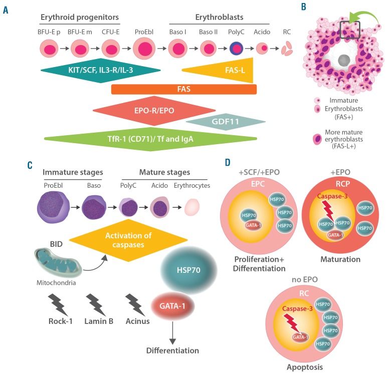
Pathological erythropoiesis with consequent anemia is a leading cause of symptomatic morbidity in internal medicine. The etiologies of anemia are complex and include reactive as well as neoplastic conditions. Clonal expansion of erythroid cells in the bone marrow may result in peripheral erythrocytosis and polycythemia but can also result in anemia when clonal cells are dysplastic and have a maturation arrest that leads to apoptosis and hinders migration, a constellation typically seen in the myelodysplastic syndromes. Rarely, clonal expansion of immature erythroid blasts results in a clinical picture resembling erythroid leukemia. Although several mechanisms underlying normal and abnormal erythropoiesis and the pathogenesis of related disorders have been deciphered in recent years, little is known about specific markers and targets through which prognosis and therapy could be improved in anemic or polycythemic patients. In order to discuss new markers, targets and novel therapeutic approaches in erythroid disorders and the related pathologies, a workshop was organized in Vienna in April 2017. The outcomes of this workshop are summarized in this review, which includes a discussion of new diagnostic and prognostic markers, the updated WHO classification, and an overview of new drugs used to stimulate or to interfere with erythropoiesis in various neoplastic and reactive conditions. The use and usefulness of established and novel erythropoiesis-stimulating agents for various indications, including myelodysplastic syndromes and other neoplasms, are also discussed.


Figures

Figure 1.
Figure 1.
Major pathways and molecules involved in the regulation of erythropoiesis. (A) Development of erythropoietic progenitor cells and erythroblasts. Early stages of erythropoietic development include primitive (p) and more mature (m) burst-forming unit erythroid cells (BFU-E) and colony-forming unit erythroid cells (CFU-E). The differentiation and maturation of these cells are regulated by broadly acting hematopoietic cytokines, including stem cell factor (SCF) and interleukin-3 (IL-3) and their receptors (R), the SCF receptor KIT and IL-3-R. Later stages are primarily regulated by erythropoietin (EPO) and EPO-R and are dependent on iron-metabolism and the interaction between the death receptor FAS and its ligand (FAS-L). Transferrin (Tf) and Tf-receptor-1 (TfR-1) are additional major regulators of erythropoiesis. Moreover, growth differentiating factor 11 (GDF11) and polymeric immunoglobulin A (IgA) are considered to be involved in the regulation of certain stages of erythropoiesis. Later stages of erythropoiesis include proerythroblasts (ProEbl), basophilic (Baso) erythroblasts (type I and II), polychromatic (PolyC) erythroblasts and acidophilic (Acido) erythroblasts, also called late erythroblasts (Barbara Bain, personal communication to MCB). FAS-L and GDF11 are involved in the final maturation stage that leads to the generation of red cells (RC). (B) Erythroid blood island: macrophage surrounded by immature erythroblasts expressing FAS and more mature erythroblasts expressing FAS-L. The cell unit (island) acts in concert to promote erythroid differentiation and red cell production and maturation (rectangle). (C) Caspase activation during terminal erythroid differentiation: BID-dependent activation of caspase occurs in mitochondria. Various caspase targets are affected, including Rock-1, Lamin B and Acinus. However, GATA-1 is protected from caspase cleavage by heat shock protein 70 (HSP70). (D) Model of terminal erythroid differentiation and apoptosis regulated by the nuclear localization of HSP70. SCF and EPO trigger the proliferation and differentiation of erythroid progenitor cells (EPC). In RC precursors (CFU-E through erythroblasts) EPO induces maturation as HSP70 translocates into the nucleus to protect GATA-1 from caspase-induced degradation. In the absence of EPO, caspase-3 induces the cleavage of GATA-1 as HSP70 cannot translocate to the nucleus, and, as a result, apoptosis occurs.
Figure 2.
Figure 2.
Immunophenotypic visualization of leukemic erythroblasts. Bone marrow sections of a patient with erythroid leukemia (acute erythroleukemia) stained with (A) Wright-Giemsa solution and antibodies against (B) glycophorin A and (C) E-cadherin. Note that virtually all leukemic erythroblasts co-express abundant amounts of glycophorin A and E-cadherin, thereby confirming the immature stage of maturation of leukemic (erythroid) cells.
Figure 3.
Figure 3.
Structure and size of erythroid islands in the bone marrow. (A) Erythroid islands in the bone marrow of a 16-year old healthy male visualized by staining for CD71. (B) Erythroid islands in the bone marrow of an 82-year old female without bone marrow neoplasm. Erythroid islands were visualized by staining against hemoglobin A. Note the decreased number and increased size of erythroid islands in the bone marrow of the older healthy control. (C) Erythroid islands in the bone marrow of a 73-year old male patient with myelodysplastic syndrome with excess of blasts (5–9% of marrow cells, MDS-EB-1) visualized by staining for CD71. In the right images of 3A, 3B and 3C, erythroid islands are evidenced by pink circles. Note the decreased number of erythroid islands in this patient. (D, left panel) Bone marrow section of a 78-year old male with myelodysplastic syndrome with excess of blasts (10–19% of marrow cells, MDS-EB-2) stained for CD71. In this patient, numerous confluent, partially disrupted and poorly separable erythroblastic islands are seen. (D, right panel) Bone marrow section of an adult patient with hemolytic anemia. Note that erythroid islands are increased, but are clearly separable and have a regular shape (contrasting with MDS). Original magnifications: A, C, D: × 125; B: × 250.

References

    1. Palis J. Primitive and definitive erythropoiesis in mammals. Front Physiol. 2014; 5:3. - PMC - PubMed
    1. Hattangadi SM, Wong P, Zhang L, Flygare J, Lodish HF. From stem cell to red cell: regulation of erythropoiesis at multiple levels by multiple proteins, RNAs, and chromatin modifications. Blood. 2011; 118(24):6258–6268. - PMC - PubMed
    1. Kuhrt D, Wojchowski DM. Emerging EPO and EPO receptor regulators and signal transducers. Blood. 2015; 125(23):3536–3541. - PMC - PubMed
    1. Liang R, Ghaffari S. Advances in understanding the mechanisms of erythropoiesis in homeostasis and disease. Br J Haematol. 2016; 174(5):661–673. - PMC - PubMed
    1. Oburoglu L, Romano M, Taylor N, Kinet S. Metabolic regulation of hematopoietic stem cell commitment and erythroid differentiation. Curr Opin Hematol. 2016; 23(3):198–205. - PubMed

Publication types