. 2018 Nov;11(6):1694-1704.
doi: 10.1038/s41385-018-0041-4. Epub 2018 Aug 3.

Cytomegalovirus promotes intestinal macrophage-mediated mucosal inflammation through induction of Smad7

Affiliations

Cytomegalovirus promotes intestinal macrophage-mediated mucosal inflammation through induction of Smad7

Evida A Dennis et al. Mucosal Immunol. 2018 Nov.

Abstract

Intestinal macrophages in healthy human mucosa are profoundly down-regulated for inflammatory responses (inflammation anergy) due to stromal TGF-β inactivation of NF-κB. Paradoxically, in cytomegalovirus (CMV) intestinal inflammatory disease, one of the most common manifestations of opportunistic CMV infection, intestinal macrophages mediate severe mucosal inflammation. Here we investigated the mechanism whereby CMV infection promotes macrophage-mediated mucosal inflammation. CMV infected primary intestinal macrophages but did not replicate in the cells or reverse established inflammation anergy. However, CMV infection of precursor blood monocytes, the source of human intestinal macrophages in adults, prevented stromal TGF-β-induced differentiation of monocytes into inflammation anergic macrophages. Mechanistically, CMV up-regulated monocyte expression of the TGF-β antagonist Smad7, blocking the ability of stromal TGF-β to inactivate NF-κB, thereby enabling MyD88 and NF-κB-dependent cytokine production. Smad7 expression also was markedly elevated in mucosal tissue from subjects with CMV colitis and declined after antiviral ganciclovir therapy. Confirming these findings, transfection of Smad7 antisense oligonucleotide into CMV-infected monocytes restored monocyte susceptibility to stromal TGF-β-induced inflammation anergy. Thus, CMV-infected monocytes that recruit to the mucosa, not resident macrophages, are the source of inflammatory macrophages in CMV mucosal disease and implicate Smad7 as a key regulator of, and potential therapeutic target for, CMV mucosal disease.


Conflict of interest statement

Conflict of Interest Statement: The authors declare no conflict of interest.

Figures

Figure 1
Figure 1. CMV infects but does not replicate in primary human intestinal macrophages
(A) Freshly isolated human intestinal macrophages were exposed to CMV at the indicated multiplicity of infection (MOI) or media (mock), and 4 days later CMV DNA was quantified by qPCR (n=3 donors). (B) CMV-infected (MOI 10) and mock-infected intestinal macrophages were analyzed on day 4 post-infection for internalized virus by fluorescence in situ hybridization (blue, DAPI-stained nuclei; green, CMV DNA; representative results, n=3 donors). Original magnification, 60x. (C) CMV-infected (MOI 5) and mock-infected intestinal macrophages were analyzed on day 4 post-infection for early CMV IE1 and UL71 gene transcripts by PCR (n=2 donors). (D) CMV-infected human foreskin fibroblasts (HFFs; MOI 0.5), human intestinal fibroblasts (Intestinal Fibs; MOI 0.5), human monocyte-derived macrophages (MDMs; MOI 1), and human intestinal macrophages (Intestinal MΦs; MOI 1) were analyzed on days 2, 4 and 6 post-infection for CMV DNA by qPCR (n=3 independent experiments). (E) CMV-infected (MOI 1) and mock-infected intestinal macrophages and CMV-infected (MOI 1) THP-1 macrophages were exposed to media (no stim), LPS (1 μg/mL) or M. tb (MOI 10) on day 6 post-infection, and CMV DNA was quantified subsequently on days 2, 3, 4, 6, 8, 12 and 14 by qPCR (representative results from 1 of 3 independent experiments). Data represent the mean +/−SEM.
Figure 2
Figure 2. CMV infection of intestinal macrophages does not promote constitutive or inducible cytokine production
(A) Primary human intestinal macrophages and (B) blood monocytes were CMV-infected (MOI of 1, 5 or 10) or mock-infected, cultured for 4 days, and then treated with media, flagellin (1 μg/mL), LPS (1 μg/mL) or LF82 (adherent invasive E. coli) (MOI 0.1) for 18 h, and the culture supernatants were harvested and analyzed for IL-6, TNF-α, or IL-1β (representative results from 1 of 3 donors; one-way ANOVA [Dunnett’s multiple comparison’s test, each group compared with mock plus stimulus]). Data represent the mean +/−SEM. *p < 0.05, **p < 0.01, ***p < 0.0001.
Figure 3
Figure 3. Pre-infection of monocytes with CMV blocks stromal down-regulation of inducible cytokine production
(A) Fresh blood monocytes were exposed to S-CM (0–1000 μg/mL) for 1 h, then flagellin or LPS (0.5 μg/mL) for 18h, and supernatants were analyzed for TNF-α (representative results from 1 of 4 donors, 3–5 S-CMs per donor); one-way ANOVA [Dunnett’s multiple comparison’s test, each group compared with 0 μg/mL S-CM plus stimulus]). (B) Monocytes were mock- or CMV-infected (MOI 5) for 24–96 h, exposed to media or S-CM (500 μg/mL) for 1 h, then flagellin (0.5 μg/mL) for 18 h, and supernatants were analyzed for IL-6 (representative results from 1 of 2 donors, 2 S-CMs per donor); one-way ANOVA [Tukey’s multiple comparison’s test]). (C) Monocytes were mock- or CMV-infected (MOI 5) for 48 h, exposed to media or S-CM (500 μg/mL) for 1 h, then flagellin or LPS (0.5 μg/mL) for 18 h. Supernatants were analyzed for TNF-α (representative results from 1 of 6 independent experiments using monocytes from 4 donors, 3–5 S-CMs per donor); Student’s t test), and total RNA was analyzed for TNF-α mRNA (representative from 1 of 3 donors, 1 S-CM per donor). (D) Monocytes were either (i) mock- or CMV-infected (MOI 5) for 96 h then exposed to CMV (500 μg/mL) for 1h or (ii) exposed to S-CM for 1 h then mock- or CMV-infected (MOI 5) for 96 h. Cells then were stimulated with LPS (1 μg/mL) for 18h, and supernatants were analyzed for IL-6 (representative from 1 of 3 donors, 1–2 S-CMs per donor); Student’s t test). Data represent means +/−SEM. *p < 0.05, **p < 0.01, ***p < 0.0001.
Figure 4
Figure 4. CMV potentiation of inducible cytokine production by monocytes is dependent on enhanced MyD88 expression
Blood monocytes were mock- or CMV-infected (MOI 5) for 3 days, exposed to media or S-CM (500 μg/mL) for 1 h, then exposed to LPS (1 μg/mL) or media for 4 h and analyzed for (A) MyD88 mRNA by qRT-PCR (representative results from 1 of 3 independent experiments using monocytes from 2 donors, 1–2 S-CMs per donor); Student’s t test) *p < 0.05 and (B) MyD88 protein by Western blot (representative results using monocytes from 2 donors, 1 S-CM per donor) with relative density analysis (composite of 3 experiments). (C) Blood monocytes were transfected with control siRNA (50 nM) or MyD88-specific siRNA (50 nM), mock- or CMV-infected (MOI 5), harvested and analyzed 2 days post-infection for MyD88 protein by Western blot to confirm MyD88 knock down (representative results from 1 of 2 donors). (D) Blood monocytes were transfected with control siRNA (50 nM) or MyD88-specific siRNA (50 nM), mock- or CMV-infected (MOI 5) for 2 days and then stimulated with LPS (0.5 μg/mL) for 18 h, after which the culture supernatants were harvested and analyzed for TNF-α (representative results from 1 of 2 donors, 2 S-CMs per donor; Student’s t test). Data represent the mean +/−SEM. *p < 0.05, **p < 0.01.
Figure 5
Figure 5. CMV blocks stromal inactivation of monocyte NF-κB p65
(A) Blood monocytes were mock- or CMV-infected (MOI 5) for 2 days, exposed to media or S-CM (500 μg/mL) for 1 h, stimulated with LPS (1 μg/mL) for 5 min and then analyzed for p-NF-κB p65 expression by flow cytometry (left and middle panels, representative donor, % expression; right panel, composite of 3 donors, mean fluorescence intensity (MFI); one-way ANOVA [Tukey’s multiple comparison’s test] *p < 0.05, **p < 0.01. (B) Blood monocytes were mock- or CMV-infected (MOI 1) for 3 days, treated with media or S-CM (100 μg/mL) for 1 h and then stimulated with LPS (1 μL/mL) for 30 min. Cells were harvested and examined by confocal microscopy, and fluorescence histograms were generated to assess nuclear translocation of NF-κB p65 (green, NF-κB p65; blue, DAPI-stained nuclei; and red, pp150+ [UL32] CMV-infected cells; representative results from 1 of 3 independent experiments). (C) Blood monocytes were mock- or CMV-infected (MOI 5) for 2 days, treated with control inhibitor (SN 50M; 100 μM) or NF-κB inhibitor (SN 50; 100 μM), with or without exposure to S-CM (500 μg/mL) for 1 h, then exposed to LPS (0.5 μg/mL) or media for 18 h. Culture supernatants were harvested and analyzed for TNF-α (representative results from 1 of 2 donors, 2 S-CMs per donor; Student’s t test). Data represent the mean +/−SEM. *p < 0.05.
Figure 6
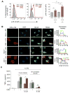
Figure 6. CMV infection of monocytes enhances Smad7 expression
Blood monocytes were mock- or CMV-infected (MOI 5) for 3 days and then analyzed for (A) Smad7 mRNA by qRT-PCR (n=3 experiments using monocytes from 3 donors; Student’s paired t test) horizontal bar corresponds to the mean. *p < 0.05; (B) Smad7 protein by Western blot (representative results from 1 of 4 independent experiments using monocytes from 2 donors) with relative density analysis (composite of 4 experiments); Student’s paired t test) horizontal bar corresponds to the mean. *p < 0.05; and (C) Smad7 localization in monocytes on days 1, 4, and 6 post-infection by immunofluorescence (red, Smad7; blue, DAPI-stained nuclei; green, UL71; Representative results from 1 of 5 experiments using monocytes from 2 donors). Original magnification, 60x. (D) Cytoplasmic localization of Smad7 was determined on day 1, 4, and 6 post-infection in mock- and CMV-infected cells. For each group, 4 different fields were chosen at random (20–50 cells per field), and the percentage of cells with cytoplasmic localization of Smad7 was calculated. Data are the mean +/− SD from analysis for each set of 4 fields. Original magnification, 20x (n=5 experiments). (E) Colon tissue specimens from a subject with ulcerative colitis and CMV infection before (upper panels) and after (lower panels) ganciclovir (GCV) therapy (n=2). Endoscopic images were obtained and biopsy consecutive sections were stained with hematoxylin & eosin and analyzed for Smad7 by immunofluorescence (red, Smad7; blue, DAPI-stained nuclei), CMV by fluorescence in situ hybridization (blue, DAPI-stained nuclei; green, CMV DNA in infected cells identified by white arrows), and CD14 (DAB). Original magnification for each 20x and insets 100x.
Figure 7
Figure 7. Smad 7 antisense oligonucleotide reverses CMV blockade of inflammation anergy
Blood monocytes were CMV-infected (MOI 5) for 1 day and then transfected with Smad7 sense or antisense oligonucleotides (2, 4 or 10 μg/mL). (A) Smad7 knock-down was confirmed 2 days later in CMV-infected cells by Western blot with relative density analysis (representative results from 1 of 2 independent experiments using monocytes from 2 donors). (B) CMV- and mock-infected monocytes were transfected 1 day post-infection with 4 μg/mL of Smad7 sense or antisense oligonucleotides, exposed to S-CM (0, 250 or 500 μg/mL) for 1 h, then flagellin (0.5 μg/mL) for 18 h, after which supernatants were analyzed for IL-6 (representative experiment, n=3, performed in duplicate with a single S-CM; one-way ANOVA [Tukey’s multiple comparison’s test]). Data represent the mean +/−SEM. For comparisons between cells transfected with antisense oligonucleotide and cells transfected with sense oligonucleotide or treated with media *p < 0.05 and **p < 0.01.

References

    1. Smith PD, Janoff EN. Complications of AIDS and other immunodeficiency states. In: Podolsky DK, Camilleri M, Fitz JG, Kalloo AN, Shanahan F, Wang TC, editors. Yamada’s Textbook of Gastroenterology. 2015. pp. 2588–2611.
    1. Janoff EN, Smith PD. Emerging concepts in gastrointestinal aspects of HIV-1 pathogenesis and management. Gastroenterology. 2001;120:607–621. - PMC - PubMed
    1. Kandiel A, Lashner B. Cytomegalovirus colitis complicating inflammatory bowel disease. Am J Gastroenterol. 2006;101:2857–2865. - PubMed
    1. Maher MM, Nassar MI. Acute cytomegalovirus infection is a risk factor in refractory and complicated inflammatory bowel disease. Dig Dis Sci. 2009;54:2456–2462. - PubMed
    1. Einhorn L, Ost A. Cytomegalovirus infection of human blood cells. J Infect Dis. 1984;149:207–214. - PubMed

Publication types