Observational Study
. 2018 Nov;39(11):4593-4610.
doi: 10.1002/hbm.24308. Epub 2018 Aug 4.

Structural network topology correlates of microstructural brain dysmaturation in term infants with congenital heart disease

Affiliations
Observational Study

Structural network topology correlates of microstructural brain dysmaturation in term infants with congenital heart disease

Vincent J Schmithorst et al. Hum Brain Mapp. 2018 Nov.

Abstract

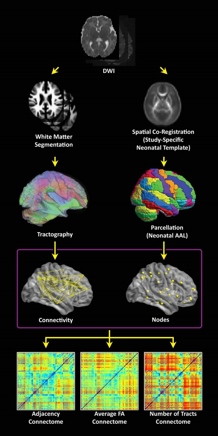
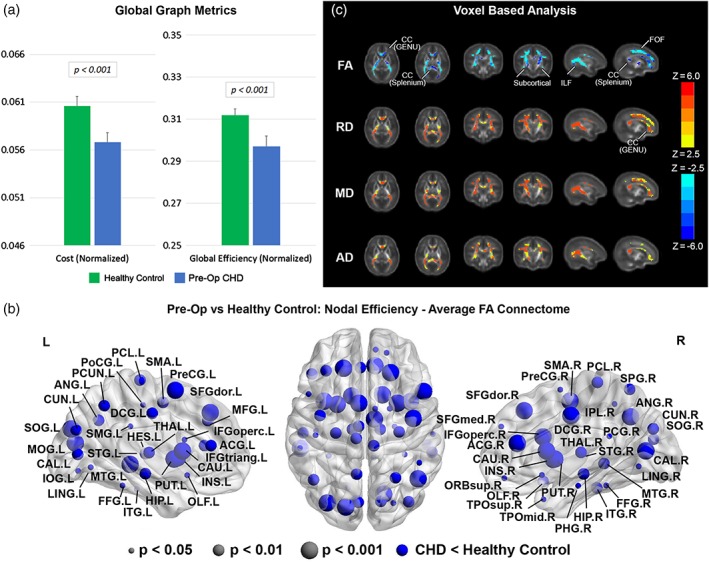
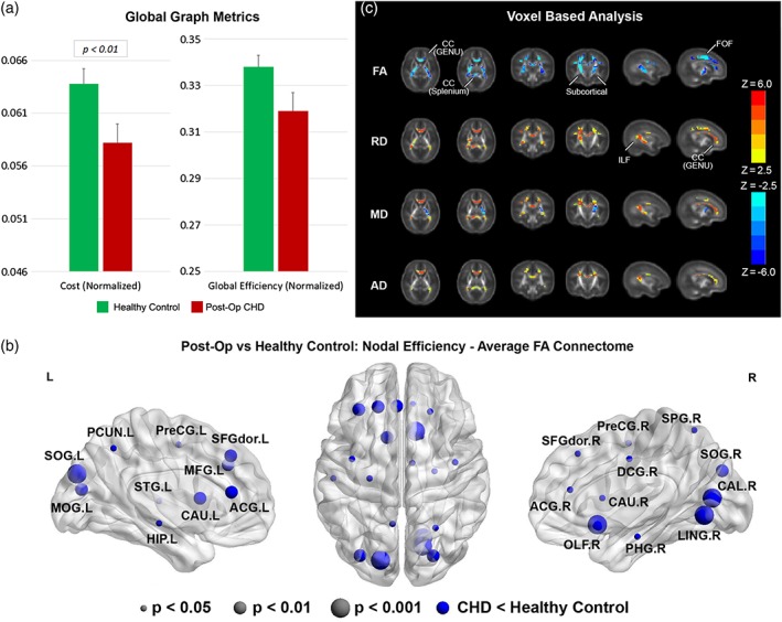
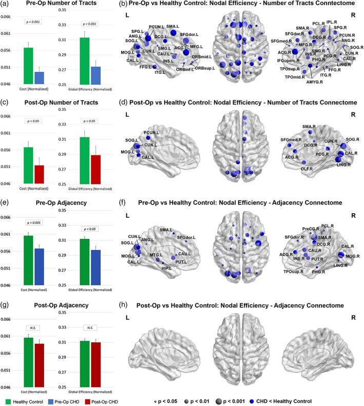
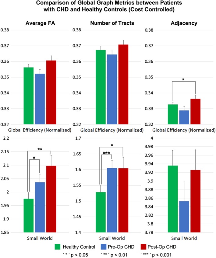
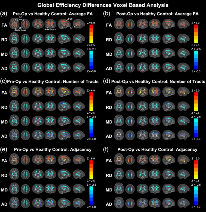
Neonates with complex congenital heart disease (CHD) demonstrate microstructural brain dysmaturation, but the relationship with structural network topology is unknown. We performed diffusion tensor imaging (DTI) in term neonates with CHD preoperatively (N = 61) and postoperatively (N = 50) compared with healthy term controls (N = 91). We used network topology (graph) analyses incorporating different weighted and unweighted approaches and subject-specific white matter segmentation to investigate structural topology differences, as well as a voxel-based analysis (VBA) to confirm the presence of microstructural dysmaturation. We demonstrate cost-dependent network inefficiencies in neonatal CHD in the pre- and postoperative period compared with controls, related to microstructural differences. Controlling for cost, we show the presence of increased small-worldness (hierarchical fiber organization) in CHD infants preoperatively, that persists in the postoperative period compared with controls, suggesting the early presence of brain reorganization. Taken together, topological microstructural dysmaturation in CHD infants is accompanied by hierarchical fiber organization during a protracted critical period of early brain development. Our methodology also provides a pipeline for quantitation of network topology changes in neonates and infants with microstructural brain dysmaturation at risk for perinatal brain injury.

Keywords: congenital heart disease; diffusion tensor MRI; graph analysis; infant.


Conflict of interest statement

The authors declare that there are no conflicts of interest or disclosures.

Figures

Figure 1
Figure 1
Flowchart depicting the data processing stream. (Right stream) the λ1 images are spatially coregistered to a neonatal T2‐weighted template (step not shown) and FA images are transformed into standard space using those transformations in order to construct a study‐specific FA neonatal template. The template is back‐transformed into native space, along with the neonatal parcellation atlas in order to define gray matter regions in native space. (Left stream) a white matter segmentation is performed in SPM8 using the FA images and neonatal GM, WM, and CSF templates. Tractography is then performed starting from each white matter voxel, which avoids the use of a strict FA threshold. (Bottom) Connectomes are computed using either: Adjacency (the matrix has 0 or 1 depending on whether there is at least one streamline connecting two regions), average FA (across all streamlines connecting two regions), or the total # of streamlines connecting two regions [Color figure can be viewed at http://wileyonlinelibrary.com]
Figure 2
Figure 2
Comparison between CHD neonates preoperatively versus normal healthy controls (average FA connectome): (a) comparison of network cost and global efficiency (values normalized to unity average graph weight); (b) comparison of nodal efficiency (all regions significant at FDR‐corrected q < 0.05); (c) comparison of DTI metrics FA, RD, MD, and AD (hot colors = CHD > controls, cold colors = CHD < controls, all regions significant at FWE‐corrected p < .05) [Color figure can be viewed at http://wileyonlinelibrary.com]
Figure 3
Figure 3
Comparison between CHD neonates postoperatively versus normal healthy controls (average FA connectome): (a) comparison of network cost and global efficiency (values normalized to unity average graph weight); (b) comparison of nodal efficiency (all regions significant at FDR‐corrected q < 0.05); (c) comparison of DTI metrics FA, RD, MD, and AD (hot colors = CHD > controls, cold colors = CHD < controls, all regions significant at FWE‐corrected p < .05) [Color figure can be viewed at http://wileyonlinelibrary.com]
Figure 4
Figure 4
Comparisons between CHD neonates pre‐ and postoperatively versus normal healthy controls: Comparison of network cost and global efficiency (values normalized to unity average graph weight) for # tracts connectome (a, c) and adjacency connectome (e, g); comparison of nodal efficiency (all regions significant at FDR‐corrected q < 0.05) for # tracts connectome (b, d) and adjacency connectome (f, h) [Color figure can be viewed at http://wileyonlinelibrary.com]
Figure 5
Figure 5
Comparison of global metrics (global efficiency, small‐worldness) between CHD neonates preoperatively, CHD neonates postoperatively, and normal healthy controls, controlling for network cost. Efficiency normalized to unity average graph weight [Color figure can be viewed at http://wileyonlinelibrary.com]
Figure 6
Figure 6
Correlation of DTI metrics (FA, RD, MD, AD) with global efficiency for preoperative CHD (a, c, e) and postoperative CHD (b, d, f) for average FA connectome (a, b), # of tracts connectome (c, d) and adjacency connectome (e, f) (hot colors = positive correlation, cold colors = negative correlation, all regions significant at FWE‐corrected p < .05) [Color figure can be viewed at http://wileyonlinelibrary.com]

References

    1. Alcauter, S. , Lin, W. , Smith, J. K. , Gilmore, J. H. , & Gao, W. (2015). Consistent anterior–posterior segregation of the insula during the first 2 years of life. Cerebral Cortex, 25(5), 1176–1187. - PMC - PubMed
    1. Ball, G. , Aljabar, P. , Zebari, S. , Tusor, N. , Arichi, T. , Merchant, N. , … Edwards, A. D. (2014). Rich‐club organization of the newborn human brain. Proceedings of the National Academy of Sciences, 111(20), 7456–7461. - PMC - PubMed
    1. Batalle, D. , Eixarch, E. , Figueras, F. , Muñoz‐Moreno, E. , Bargallo, N. , Illa, M. , … Gratacos, E. (2012). Altered small‐world topology of structural brain networks in infants with intrauterine growth restriction and its association with later neurodevelopmental outcome. NeuroImage, 60(2), 1352–1366. - PubMed
    1. Beca, J. , Gunn, J. K. , Coleman, L. , Hope, A. , Reed, P. W. , Hunt, R. W. , … Shekerdemian, L. S. (2013). New white matter brain injury after infant heart surgery is associated with diagnostic group and the use of circulatory arrest. Circulation, 127(9), 971–979. - PubMed
    1. Bellinger, D. C. , Watson, C. G. , Rivkin, M. J. , Robertson, R. L. , Roberts, A. E. , Stopp, C. , … Wypij, D. (2015). Neuropsychological status and structural brain imaging in adolescents with single ventricle who underwent the Fontan procedure. Journal of the American Heart Association, 4(12), e002302. - PMC - PubMed

Publication types