. 2019 Jan;77(1):42-53.
doi: 10.1016/j.joms.2018.06.175. Epub 2018 Jul 10.

Effect of Measurement Technique on TMJ Mandibular Condyle and Articular Disc Morphometry: CBCT, MRI, and Physical Measurements

Affiliations

Effect of Measurement Technique on TMJ Mandibular Condyle and Articular Disc Morphometry: CBCT, MRI, and Physical Measurements

Matthew C Coombs et al. J Oral Maxillofac Surg. 2019 Jan.

Abstract

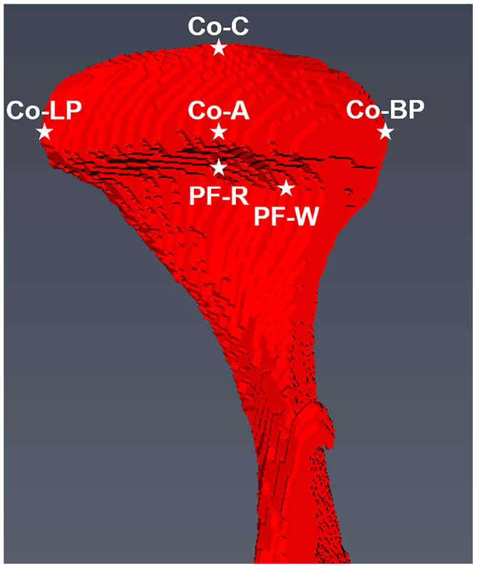
Purpose: Accurate description of the temporomandibular size and shape (morphometry) is critical for clinical diagnosis and surgical planning and the design and development of regenerative scaffolds and prosthetic devices and to model the temporomandibular loading environment. The study objective was to determine the 3-dimensional morphometry of the temporomandibular joint (TMJ) condyle and articular disc using cone-beam computed tomography (CBCT), magnetic resonance imaging (MRI), and physical measurements of the same joints using a repeated measures design and to determine the effect of the measurement technique on temporomandibular size and shape.

Materials and methods: Human cadaveric heads underwent a multistep protocol to acquire physiologically meaningful measurements of the condyle and disc. The heads first underwent CBCT scanning, and solid models were automatically generated. The superficial soft tissues were dissected, and intact TMJs were excised and underwent MRI scanning, with solid models generated after manual segmentation. After MRI, the intact joints were dissected, and physical measurements of the condyle and articular disc were performed. The CBCT-based model measurements, MRI-based model measurements, and physical measurements were standardized, and a repeated measures study design was used to determine the effect of the measurement technique on the morphometric parameters.

Results: Multivariate general linear mixed effects models showed significant effects for measurement technique for condylar morphometric outcomes (P < .001) and articular disc morphometric outcomes (P < .001). The physical measurements after dissection were larger than either the CBCT-based or MRI-based measurements. Differences in imaging-based morphometric parameters followed a complex relationship between imaging modality resolution and contrast between tissue types.

Conclusions: Physical measurements after dissection are still considered the reference standard. However, owing to their inaccessibility in vivo, understanding how the imaging technique affects the temporomandibular size and shape is critical toward the development of high-fidelity solid models to be used in the design and development of regenerative scaffolds, surgical planning, prosthetic devices, and anatomic investigations.


Figures

Figure 1.
Figure 1.
CBCT scans of donor cadaveric heads used for condylar reconstructions in the saggital and coronal planes (A). MRI scans of excised temporomandibular joints used for condylar and articular disc reconstructions in the saggital and coronal planes (B). Unsmoothed reconstruction of the mandible from CBCT scans, used to determine condylar morphometry in the saggital and coronal planes (C). Unsmoothed reconstruction of the mandible and articular disc from MRI scans, used to determine condylar and disc morphometry in the saggital and coronal planes (D).
Figure 1.
Figure 1.
CBCT scans of donor cadaveric heads used for condylar reconstructions in the saggital and coronal planes (A). MRI scans of excised temporomandibular joints used for condylar and articular disc reconstructions in the saggital and coronal planes (B). Unsmoothed reconstruction of the mandible from CBCT scans, used to determine condylar morphometry in the saggital and coronal planes (C). Unsmoothed reconstruction of the mandible and articular disc from MRI scans, used to determine condylar and disc morphometry in the saggital and coronal planes (D).
Figure 2.
Figure 2.
Illustrations of the condylar morphometric parameters in the axial plane (A) and coronal plane (B), with illustrations of the disc morphometric parameters in the axial plane (C) and orthogonal view (D). Temporomandibular joint morphometric parameters were standardized between the CBCT-based model measurements, MRI-based model measurements and physical measurements. Condylar morphometric outcomes were: major axis length, defined as the distance from Co-LP to Co-BP; minor axis length, defined as the distance from Co-A to Co-P; condylar height, defined as the distance from PF-R to Co-C; lingual length, defined as the distance from PF-W to Co-LP; buccal length, defined as the distance from PF-W to Co-BP. Articular disc morphometric outcomes were: major axis length, defined as the distance from D-LP to D-BP; minor axis length, defined as the distance from D-A to D-P; anterior band thickness, defined as the distance from the superior surface of the disc to the inferior surface located at D-A (shown only); posterior band thickness, defined as the distance from the superior surface of the disc to the inferior surface located at D-P; medial aspect thickness, defined as the distance from the superior surface of the disc to the inferior surface located at D-LP; lateral aspect thickness, defined as the distance from the superior surface of the disc to the inferior surface located at D-BP; intermediate zone thickness, defined as the distance from the superior surface of the disc to the inferior surface of the disc at the disc center (D-IZ). Mandibular condyle and articular disc morphometric measurements were all made on unsmoothed 3D models, to limit the effects of shrinkage relative to their natural physical dimensions during smoothing processes.
Figure 2.
Figure 2.
Illustrations of the condylar morphometric parameters in the axial plane (A) and coronal plane (B), with illustrations of the disc morphometric parameters in the axial plane (C) and orthogonal view (D). Temporomandibular joint morphometric parameters were standardized between the CBCT-based model measurements, MRI-based model measurements and physical measurements. Condylar morphometric outcomes were: major axis length, defined as the distance from Co-LP to Co-BP; minor axis length, defined as the distance from Co-A to Co-P; condylar height, defined as the distance from PF-R to Co-C; lingual length, defined as the distance from PF-W to Co-LP; buccal length, defined as the distance from PF-W to Co-BP. Articular disc morphometric outcomes were: major axis length, defined as the distance from D-LP to D-BP; minor axis length, defined as the distance from D-A to D-P; anterior band thickness, defined as the distance from the superior surface of the disc to the inferior surface located at D-A (shown only); posterior band thickness, defined as the distance from the superior surface of the disc to the inferior surface located at D-P; medial aspect thickness, defined as the distance from the superior surface of the disc to the inferior surface located at D-LP; lateral aspect thickness, defined as the distance from the superior surface of the disc to the inferior surface located at D-BP; intermediate zone thickness, defined as the distance from the superior surface of the disc to the inferior surface of the disc at the disc center (D-IZ). Mandibular condyle and articular disc morphometric measurements were all made on unsmoothed 3D models, to limit the effects of shrinkage relative to their natural physical dimensions during smoothing processes.
Figure 3.
Figure 3.
Significant differences in condylar morphometry between measurement techniques were determined (p<001), with significant differences for condylary minor axis length (p=0.002), condylar height (p<0.001), condylar buccal length (p<0.001), and condylar lingual length (p<0.001). Individual pairwise contrasts in condylar morphometric outcomes between measurement techniques were determined: *, p<0.05; **, p<0.01; ***, p<0.001.
Figure 4.
Figure 4.
Significant differences in articular disc morphometry between measurement techniques were determined (p<001). Individual pairwise contrasts in articular disc morphometric outcomes between measurement techniques were determined: *, p<0.05; **, p<0.01; ***, p<0.001.

References

    1. Haskin CL, Milam SB, Cameron IL: Pathogenesis of degenerative joint disease in the human temporomandibular joint. Crit Rev Oral Biol Med 6:248, 1995 - PubMed
    1. Zizelmann C, Bucher P, Rohner D, Gellrich NC, Kokemueller H, Hammer B: Virtual restoration of anatomic jaw relationship to obtain a precise 3D model for total joint prosthesis construction for treatment of TMJ ankylosis with open bite. Int J Oral Maxillofac Surg 39:1012, 2010 - PubMed
    1. Matsumoto K, Ishiduka T, Yamada H, Yonehara Y, Arai Y, Honda K: Clinical use of three-dimensional models of the temporomandibular joint established by rapid prototyping based on cone-beam computed tomography imaging data. Oral Radiology 30:98, 2014
    1. Legemate K, Tarafder S, Jun Y, Lee CH: Engineering Human TMJ Discs with Protein- Releasing 3D-Printed Scaffolds. J Dent Res 95:800, 2016 - PubMed
    1. Brown BN, Chung WL, Almarza AJ, Pavlick MD, Reppas SN, Ochs MW, Russell AJ, Badylak SF: Inductive, scaffold-based, regenerative medicine approach to reconstruction of the temporomandibular joint disk. J Oral Maxillofac Surg 70:2656, 2012 - PMC - PubMed

MeSH terms

LinkOut - more resources