. 2018 Oct:332:58-76.
doi: 10.1016/j.cellimm.2018.07.008. Epub 2018 Jul 19.

Similarities and differences in surface receptor expression by THP-1 monocytes and differentiated macrophages polarized using seven different conditioning regimens

Affiliations

Similarities and differences in surface receptor expression by THP-1 monocytes and differentiated macrophages polarized using seven different conditioning regimens

Megan A Forrester et al. Cell Immunol. 2018 Oct.

Abstract

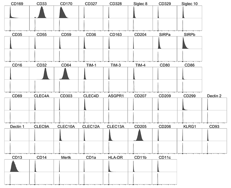
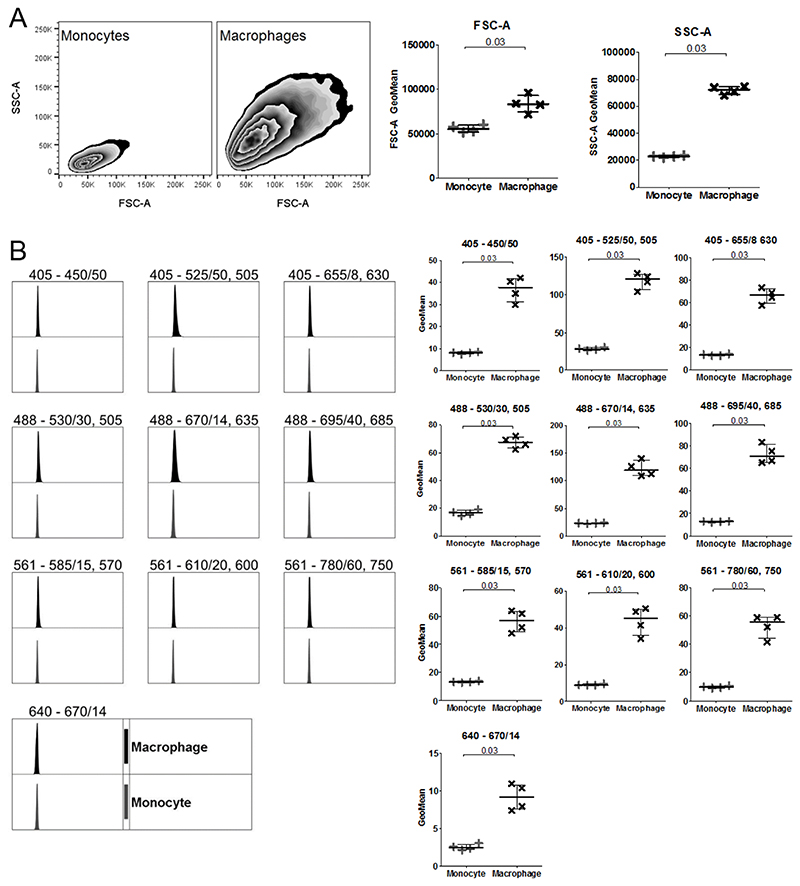
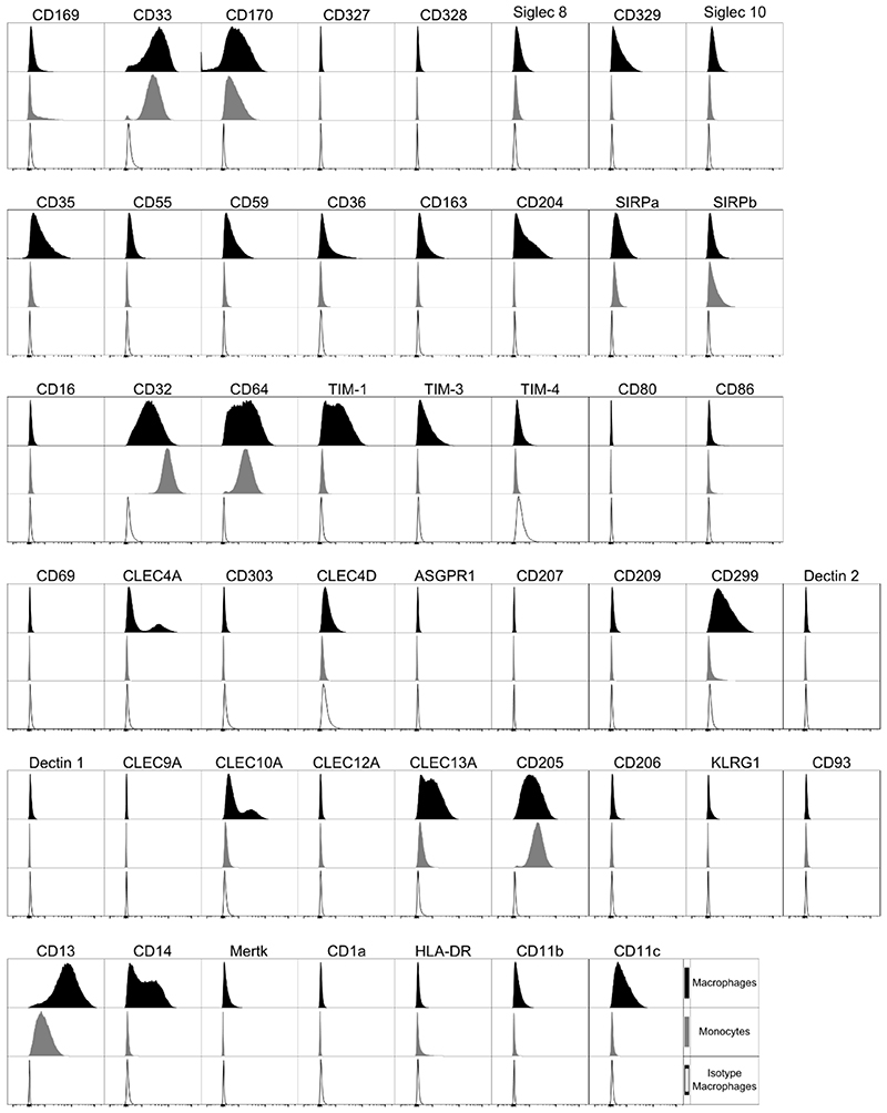
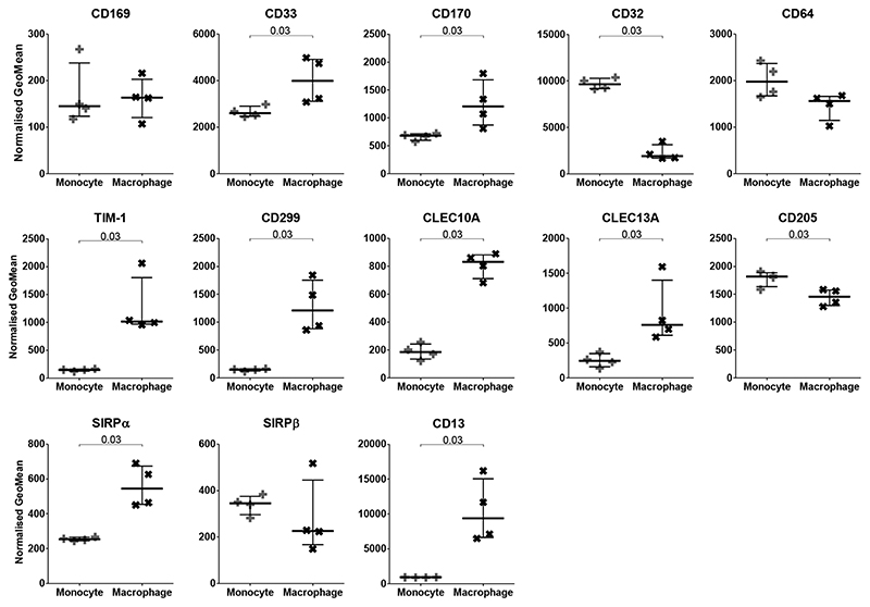
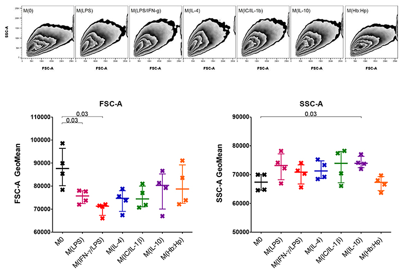
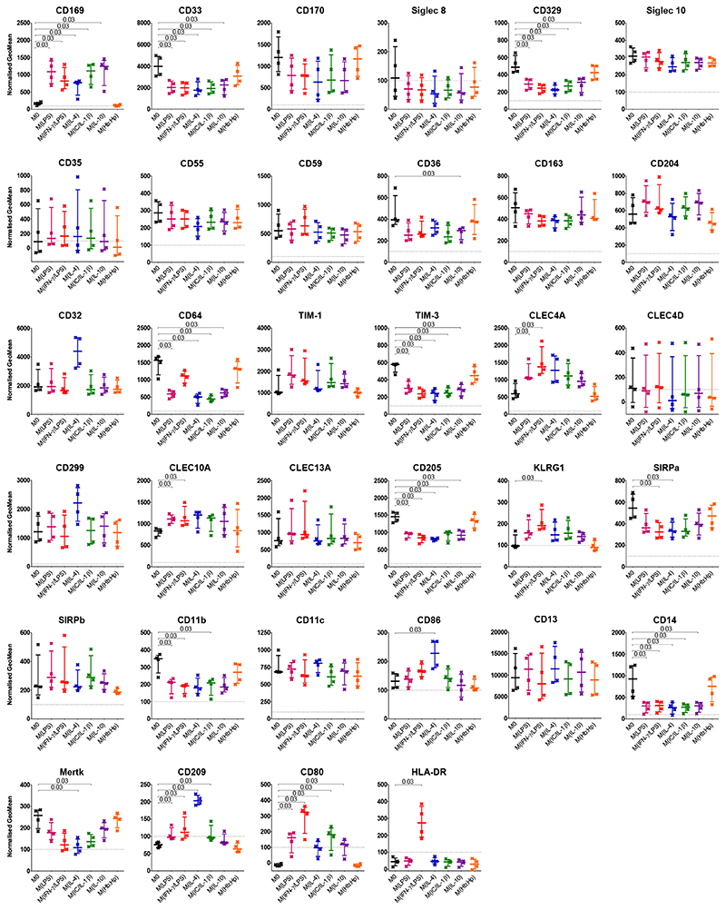
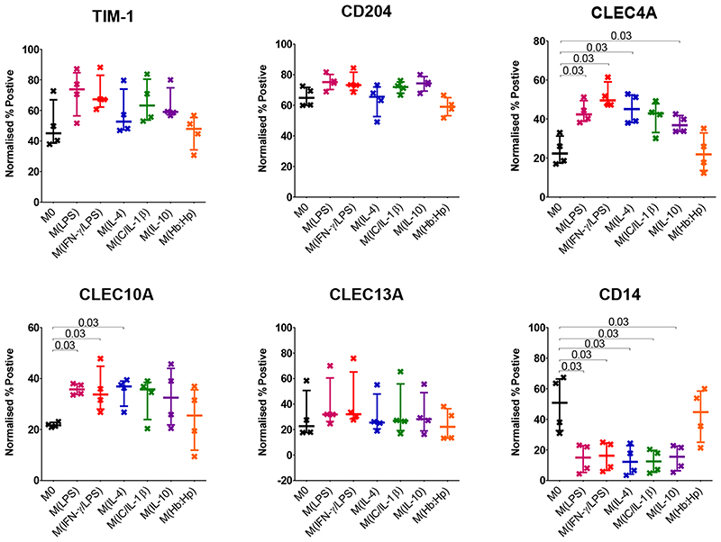
Macrophages are key in orchestrating immune responses to micro-environmental stimuli, sensed by a complex set of surface receptors. The human cell line THP-1 has a monocytic phenotype, including the ability to differentiate into macrophages, providing a tractable, standardised surrogate for human monocyte-derived macrophages. Here we assessed the expression of 49 surface markers including Fc, complement, C-type lectin and scavenger receptors; TIMs; Siglecs; and co-stimulatory molecules by flow cytometry on both THP-1 monocytes and macrophages and following macrophage activation with seven standard conditioning/polarizing stimuli. Of the 34 surface markers detected on macrophages, 18 altered expression levels on activation. From these, expression of 9 surface markers were consistently altered by all conditioning regimens, while 9 were specific to individual polarizing stimuli. This study provides a resource for the study of macrophages and highlights that macrophage polarization states share much in common and the differences do not easily fit a simple classification system.

Keywords: Cell surface; Flow cytometry; Macrophage; Monocyte; Polarization; THP-1.


Conflict of interest statement

Disclosure of Conflicts of Interest

The authors declare no competing interests.

Figures

Figure 1
Figure 1. Cell Marker Expression on THP-1 Monocytes.
Filled histogram target antibody stained, open histogram matched isotype control. Histograms are representative of n = 4.
Figure 2
Figure 2. Comparison of flow cytometric cell features between THP-1 Monocytes and Macrophages.
A Size (FSC-A) and granularity (SSC-A) of monocytes and macrophages. B Unstained monocytes (grey; +) and macrophages (black; x) shows autofluorescence within each cytometer channel. Histograms and zebra plots are representative of n = 4. GeoMean was normalised by subtracting the unstained value from the isotype control. Median with IQR; Mann-Whitney tests.
Figure 3
Figure 3. Cell Marker Expression on THP-1 Macrophages.
PMA differentiated THP-1 macrophages. Black filled histogram target antibody stained macrophages, grey filled histogram target antibody stained monocytes, open histogram matched isotype control macrophages. Histograms are representative of n = 4.
Figure 4
Figure 4. Comparison of Cell Marker Expression between THP-1 Monocytes and Macrophages.
Normalized GeoMeans of markers present on both monocytes (+) and macrophages (x) compared. GeoMean was normalised by subtracting the matched isotype control value. N=4; Median with IQR; Mann-Whitney tests.
Figure 5
Figure 5. Cell size and granularity of THP-1 Macrophages after 24 Hours Phenotype Biasing.
Size (FSC-A) and granularity (SSC-A) of phenotype biased macrophages, zebra plots representative of n = 4. Median with IQR; Mann-Whitney tests.
Figure 6
Figure 6. Cell Marker Expression on THP-1 Macrophages after 24 Hours Phenotype Biasing.
PMA differentiated THP-1 macrophages after phenotype biasing for 24 hours. No stimuli (M0) black, LPS red, LPS and IFN-γ pink, IL-4 blue, IL-1β and immune complex green, IL-10 purple, hemoglobin:haptoglobin orange. GeoMean was normalised by subtracting the matched isotype control value. N=4; Median with IQR; Mann-Whitney tests.
Figure 7
Figure 7. Percentage Positive Cell Marker Expression on THP-1 Macrophages after 24 Hours Phenotype Biasing.
PMA differentiated THP-1 macrophages after phenotype biasing for 24 hours. No stimuli (M0) black, LPS red, LPS and IFN-γ pink, IL-4 blue, IL-1β and immune complex green, IL-10 purple, hemoglobin:haptoglobin orange. Percentage positive determined by isotype control then normalised by subtracting the matched isotype control value. N=4; Median with IQR; Mann-Whitney tests.

References

    1. Murray PJ, Wynn TA. Protective and pathogenic functions of macrophage subsets. Nat Rev Immunol. 2011;11:723–737. - PMC - PubMed
    1. Martinez FO, Sica A, Mantovani A, Locati M. Macrophage activation and polarization, Front. Biosci. 2008:453–461. - PubMed
    1. Martinez FO, Gordon S. The M1 and M2 paradigm of macrophage activation: time for reassessment. F1000Prime Rep. 2014:13–13.:eCollection 2014. - PMC - PubMed
    1. Boyle JJ, Harrington HA, Piper E, Elderfield K, Stark J, Landis RC, Haskard DO. Coronary intraplaque hemorrhage evokes a novel atheroprotective macrophage phenotype. Am J Pathol. 2009;3:1097–1108. - PMC - PubMed
    1. Boyle JJ, Johns M, Kampfer T, Nguyen AT, Game L, Schaer DJ, Mason JC, Haskard DO. Activating transcription factor 1 directs Mhem atheroprotective macrophages through coordinated iron handling and foam cell protection. Circ Res. 2012;1:20–33. - PubMed

Publication types

LinkOut - more resources