. 2018 Aug;119(5):580-590.
doi: 10.1038/s41416-018-0200-0. Epub 2018 Aug 6.

Identification of beta-arrestin-1 as a diagnostic biomarker in lung cancer

Affiliations

Identification of beta-arrestin-1 as a diagnostic biomarker in lung cancer

Victoria El-Khoury et al. Br J Cancer. 2018 Aug.

Abstract

Background: Distinguishing lung adenocarcinoma (ADC) from squamous cell carcinoma (SCC) has a tremendous therapeutic implication. Sometimes, the commonly used immunohistochemistry (IHC) markers fail to discriminate between them, urging for the identification of new diagnostic biomarkers.

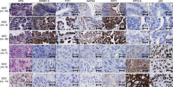
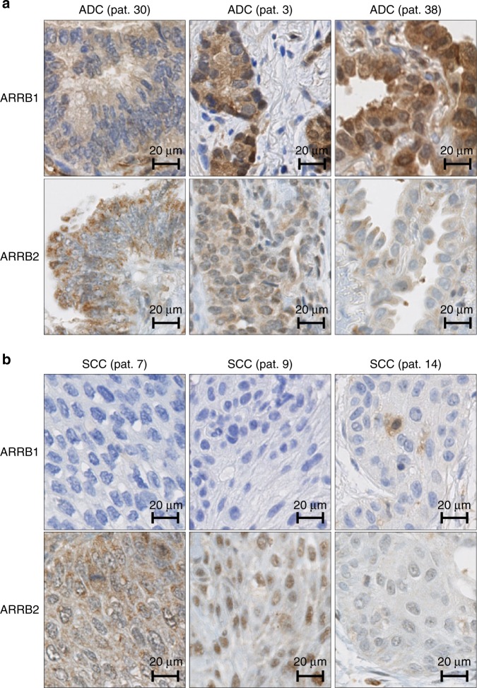
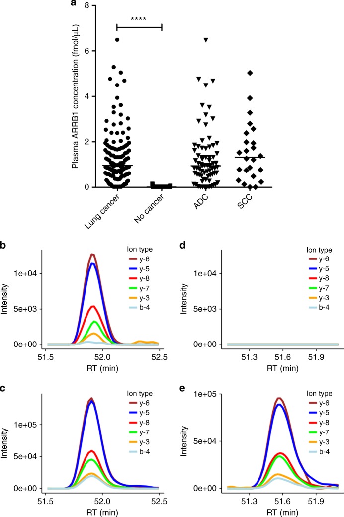
Methods: We performed IHC on tissue microarrays from two cohorts of lung cancer patients to analyse the expression of beta-arrestin-1, beta-arrestin-2 and clinically used diagnostic markers in ADC and SCC samples. Logistic regression models were applied for tumour subtype prediction. Parallel reaction monitoring (PRM)-based mass spectrometry was used to quantify beta-arrestin-1 in plasma from cancer patients and healthy donors.

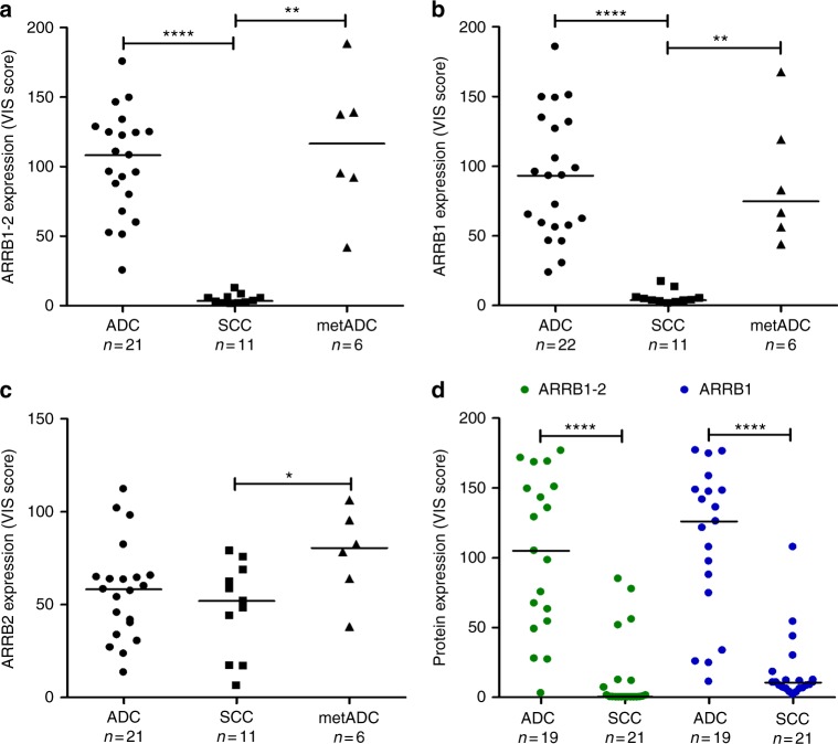
Results: Beta-arrestin-1 expression was significantly higher in ADC versus SCC samples. Beta-arrestin-1 displayed high sensitivity, specificity and negative predictive value. Its usefulness in an IHC panel was also shown. Plasma beta-arrestin-1 levels were considerably higher in lung cancer patients than in healthy donors and were higher in patients who later experienced a progressive disease than in patients showing complete/partial response following EGFR inhibitor therapy.

Conclusions: Our data identify beta-arrestin-1 as a diagnostic marker to differentiate ADC from SCC and indicate its potential as a plasma biomarker for non-invasive diagnosis of lung cancer. Its utility to predict response to EGFR inhibitors is yet to be confirmed.


Conflict of interest statement

The authors declare no competing interests.

Figures

Fig. 1
Fig. 1
Beta-arrestin-1 and 2 protein expression in lung ADC and SCC samples in the in-house and the amsbio TMAs. Scatter plots represent quantitative analysis (VIS score) of IHC results in sections of ac the in-house TMA and of d the amsbio TMA. Sections of the in-house TMA were incubated with an antibody that (a) recognises both beta-arrestin-1 and 2 (ARRB1-2), or specifically detects either (b) beta-arrestin-1 (ARRB1) or c beta-arrestin-2 (ARRB2). d Sections of the amsbio TMA were incubated with the anti-beta-arrestin-1-2 antibody or with the specific anti-beta-arrestin-1 antibody and “n” indicates the number of subjects in each group. Data points and their median are shown. ****P < 0.0001 **P < 0.01 and *P < 0.05 using Mann–Whitney rank sum test
Fig. 2
Fig. 2
Representative images of HPS and IHC stainings in lung ADC and SCC samples. Automated IHC was performed on sections of the in-house TMA using antibodies that recognise beta-arrestin-1-2 (ARRB1-2), the commonly used ADC markers (TTF1, NAPSA, KRT7) and SCC markers (KRT5-6 and p63). DAB-based visualisation was used to assess protein expression. For each sample, a haematoxylin/phloxine/saffron (HPS)-stained slide is shown
Fig. 3
Fig. 3
Representative images of beta-arrestin-1 (ARRB1) and beta-arrestin-2 (ARRB2) IHC staining in lung ADC and SCC samples. Automated IHC was performed on sections from the in-house TMA using antibodies that recognise specifically beta-arrestin-1 or beta-arrestin-2. DAB-based visualisation was used to assess protein expression. IHC images in a three ADC samples and b three SCC samples are shown. Results from the same patient samples as those represented in Fig. 2 are displayed here
Fig. 4
Fig. 4
Plasma levels of beta-arrestin-1 (ARRB1) in lung cancer patients and healthy donors. a Scatter plots of plasma beta-arrestin-1 concentration obtained from lung cancer patients (n = 128) and healthy volunteers (n = 93) using the PRM assay targeting EDLDVLGLTFR. Data points and their median are shown. PRM data from the lung cancer patients were further divided into ADC (n = 72) and SCC (n = 24) groups in the same graph. PRM readout of beta-arrestin-1 measured in one of the lung cancer patient samples is shown in b with the traces of detected product ions of the peptide EDLDVLGLTFR compared to the ones of internal standard peptide shown in c. PRM readout of beta-arrestin-1 measured in one of the healthy volunteer samples is shown in d indicating no signal was detected compared to the internal standard shown in e. ****P < 0.0001 using Mann–Whitney rank sum test

References

    1. Bennett CW, Berchem G, Kim YJ, El-Khoury V. Cell-free DNA and next-generation sequencing in the service of personalized medicine for lung cancer. Oncotarget. 2016;7:71013–71035. doi: 10.18632/oncotarget.11717. - DOI - PMC - PubMed
    1. Zhu QG, Zhang SM, Ding XX, He B, Zhang HQ. Driver genes in non-small cell lung cancer: characteristics, detection methods, and targeted therapies. Oncotarget. 2017;8:57680–57692. - PMC - PubMed
    1. Ring BZ, et al. A novel five-antibody immunohistochemical test for subclassification of lung carcinoma. Mod. Pathol. 2009;22:1032–1043. doi: 10.1038/modpathol.2009.60. - DOI - PubMed
    1. Takamochi K, et al. Novel biomarkers that assist in accurate discrimination of squamous cell carcinoma from adenocarcinoma of the lung. BMC Cancer. 2016;16:760. doi: 10.1186/s12885-016-2792-1. - DOI - PMC - PubMed
    1. Chiu CH, Chou TY, Chiang CL, Tsai CM. Should EGFR mutations be tested in advanced lung squamous cell carcinomas to guide frontline treatment? Cancer Chemother. Pharmacol. 2014;74:661–665. doi: 10.1007/s00280-014-2536-3. - DOI - PubMed

Publication types

MeSH terms