Case Reports
. 2018 Jul 27;10(7):523-529.
doi: 10.4254/wjh.v10.i7.523.

Hepatectomy for gallbladder-cancer with unclassified anomaly of right-sided ligamentum teres: A case report and review of the literature

Affiliations
Case Reports

Hepatectomy for gallbladder-cancer with unclassified anomaly of right-sided ligamentum teres: A case report and review of the literature

Toru Goto et al. World J Hepatol. .

Abstract

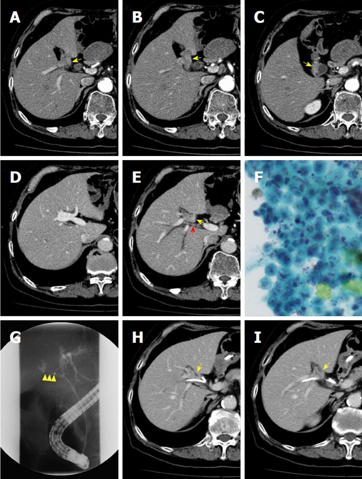
Right-sided ligamentum teres (RSLT) is a congenital anomaly in which the right umbilical ligament becomes dominant and anomalous ramifications of the hepatic vessels and biliary system are present. A male patient in his 70s was diagnosed with advanced gallbladder cancer directly infiltrating the right hepatic duct (RHD), together with RSLT. Preoperative three-dimensional simulation of the liver based on multiple detector computed tomography images after cholangiography revealed ramifications of all segmental portal veins from the portal trunk and discordance of the arterial and biliary branching patterns of segment 8. Fusion analysis of the biliary architecture and segmental volumetry showed that the RHD drained segments 1r, 5, 6, and 7. We successfully performed a modified right-sided hepatectomy sparing segment 8 (i.e., resection of the RHD drainage territory), with negative surgical margins. This report is the first to describe major hepatectomy for advanced gallbladder cancer with RSLT.

Keywords: Anomaly of the portal vein; Gallbladder cancer; Hepatectomy; Preoperative liver simulation; Right-sided ligamentum teres.


Conflict of interest statement

Conflict-of-interest statement: The authors declare that no conflicts of interest are associated with this manuscript.

Figures

Figure 1
Figure 1
Contrast-enhanced computed tomography, cytological examination of bile, and endoscopic retrograde cholangiography. A, B and C: The highly atrophied gallbladder (yellow arrow) had tumor-like localized wall thickness enhanced by contrast medium; D: The cul-de-sac of the right-sided umbilical portion is shown; E: The gallbladder (yellow arrow) is located on the left-sided liver bed. Direct infiltration of the tumor into the right hepatic artery (red arrow) is not evident; F: Cytological examination of bile obtained from endoscopic retrograde gallbladder drainage demonstrated atypical cells with a high nucleo-cytoplasmic ratio, strongly suggesting adenocarcinoma; G: Endoscopic retrograde cholangiography shows severe stenosis of the right hepatic duct (yellow arrowhead); H and I: The gallbladder tumor (yellow arrow) directly spreads to the nearby right hepatic duct where an endoscopic nasobiliary tube was placed.
Figure 2
Figure 2
Preoperative simulation. A: An “all-in-one” simulation image. The intrahepatic vasculature was reconstructed at the 4th order division level (red: hepatic artery, pink: portal vein, green: bile duct, yellow: Gallbladder, blue: hepatic vein); B and C: Simulated segmentation based on the portal venous flow. Couinaud’s definition was referred to for the naming of each segment; D: All segmental portal branches were ramified from the portal trunk. Interestingly, a common trunk of P5 and P6 was present; E: Hepatic arterial ramification without anomalous anatomy; F: The bile duct of segment 8 (B8) was ramified from the left hepatic duct, not from the right hepatic duct. RHD: Right hepatic duct; LHD: Left hepatic duct.
Figure 3
Figure 3
Simulation of modified right-sided hepatectomy. The resection area corresponded to the drainage territory of the right hepatic duct (segments 1r, 5, 6, and 7, blue area).
Figure 4
Figure 4
Hepatic hilar view during the operation. The gallbladder was located at the left side of the right-sided ligamentum teres (RSLT).
Figure 5
Figure 5
Demarcation line after dividing all arterial and portal branches of segments 1r, 5, 6, and 7.
Figure 6
Figure 6
Final view of the hepatic hilum after hepatectomy. The portal and hepatic arterial branches of segment 8 were preserved. A common trunk of P5 and P6 was detected during the operation, which was confirmed by ischemic changes in S5 and S6 after clamping the target branch.
Figure 7
Figure 7
Final view after hepaticojejunostomy. The ligamentum teres originated from the main portal trunk and ran between S4-6 and S4-7.
Figure 8
Figure 8
Pathological examination. Pathological examination shows well-differentiated tubular adenocarcinoma of the gallbladder (T) with direct invasion of the liver parenchyma and the right hepatic duct (RHD: white arrow). The tumor cell invaded to the RHD wall, but not into the lumen (yellow arrowhead) (hematoxylin eosin saffron, original magnification × 20).

References

    1. Nagai M, Kubota K, Kawasaki S, Takayama T, BandaiY, Makuuchi M. Are left-sided gallbladders really located on the left side? Ann Surg. 1997;225:274–280. - PMC - PubMed
    1. Maetani Y, Itoh K, Kojima N, Tabuchi T, Shibata T, Asonuma K, Tanaka K, Konishi J. Portal vein anomaly associated with deviation of the ligamentum teres to the right and malposition of the gallbladder. Radiology. 1998;207:723–728. - PubMed
    1. Shindoh J, Akahane M, Satou S, Aoki T, Beck Y, Hasegawa K, Sugawara Y, Ohtomo K, Kokudo N. Vascular architecture in anomalous right-sided ligamentum teres: three-dimensional analyses in 35 patients. HPB (Oxford) 2012;14:32–41. - PMC - PubMed
    1. Nishitai R, Shindoh J, Yamaoka T, Akahane M, Kokudo N, Manaka D. Biliary architecture of livers exhibiting right-sided ligamentum teres: an indication for preoperative cholangiography prior to major hepatectomy. HPB (Oxford) 2016;18:929–935. - PMC - PubMed
    1. Kawai K, Koizumi M, Honma S, Tokiyoshi A, Kodama K. Right ligamentum teres joining to the right branch of the portal vein. Anat Sci Int. 2008;83:49–54. - PubMed

Publication types

LinkOut - more resources