. 2018 Jun 12;9(27):5929-5936.
doi: 10.1039/c8sc01233d. eCollection 2018 Jul 21.

Pyrophosphorylation via selective phosphoprotein derivatization

Affiliations

Pyrophosphorylation via selective phosphoprotein derivatization

Alan M Marmelstein et al. Chem Sci. .

Abstract

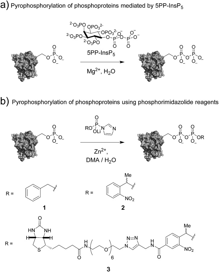
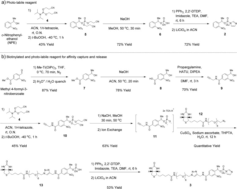
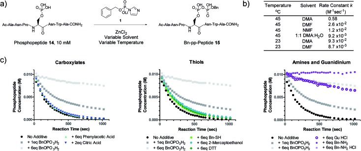
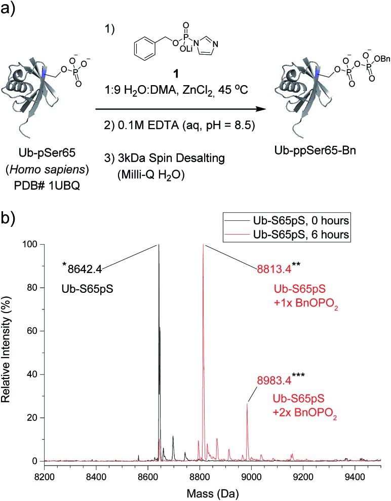
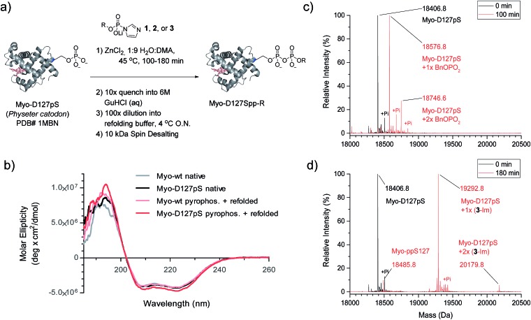
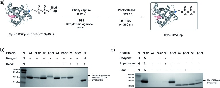
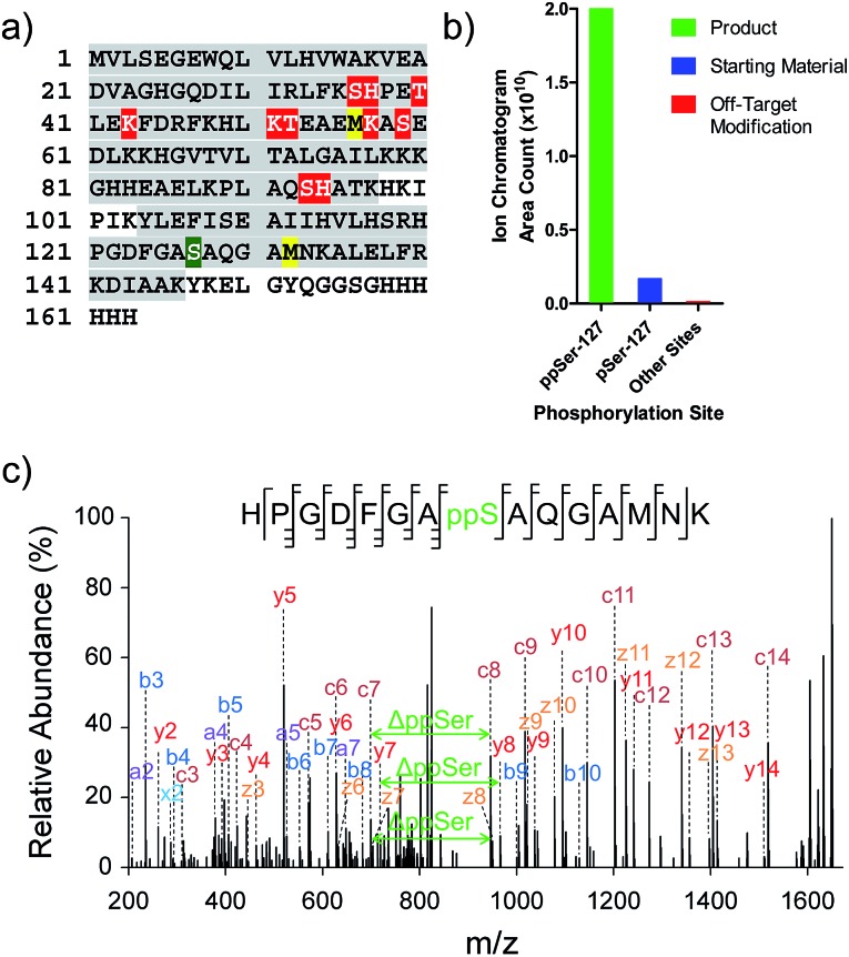
An important step in elucidating the function of protein post-translational modifications (PTMs) is gaining access to site-specifically modified, homogeneous samples for biochemical characterization. Protein pyrophosphorylation is a poorly characterized PTM, and here a chemical approach to obtain pyrophosphoproteins is reported. Photo-labile phosphorimidazolide reagents were developed for selective pyrophosphorylation, affinity-capture, and release of pyrophosphoproteins. Kinetic analysis of the reaction revealed rate constants between 9.2 × 10⁻³ to 0.58 M-1 s-1, as well as a striking proclivity of the phosphorimidazolides to preferentially react with phosphate monoesters over other nucleophilic side chains. Besides enabling the characterization of pyrophosphorylation on protein function, this work highlights the utility of phosphoryl groups as handles for selective protein modification for a variety of applications, such as phosphoprotein bioconjugation and enrichment.


Figures

Fig. 1
Fig. 1. (a) Protein pyrophosphorylation by 5PP-InsP5, and (b) chemical pyrophosphorylation using phosphorimidazolide reagents.
Scheme 1
Scheme 1. Synthesis of phosphorimidazolide reagents.
Fig. 2
Fig. 2. Kinetic characterization of pyrophosphorylation reactions. (a) Reaction of model phosphopeptide 14 with P-imidazolide 1. (b) Tabulated pseudo-first order kinetic data. Conditions: 100 mM P-imidazolide 1, 10 mM phosphopeptide 14, 267 mM ZnCl2. (c) Reaction progress kinetic data for the reaction of phosphopeptide 14 with reagent 1 in the presence of various additives. Data points represent averages from three replicate reactions (except for runs with added 2-mercaptoethanol, DTT, and guanidine HCl). Error bars are omitted for clarity but are depicted in Fig. S7. Processed RPKA data is shown in Fig. S8. Conditions: 10 mM peptide 4, 30 mM P-imidazolide 1, 80 mM ZnCl2, in 1 : 9 H2O : DMA at 45 °C.
Fig. 3
Fig. 3. Pyrophosphorylation and characterization of ubiquitin-S65pS. (a) Pyrophosphorylation procedure for Ub-S65pS. 306 mM ZnCl2, 61.2 mM reagent 1 and 29 μM protein. (b) ESI-MS of Ub-S65pS before pyrophosphorylation (black) and after 6 hours under pyrophosphorylation reaction conditions (red). Mass error of 10 ppm; mass resolution of 0.1 Da.
Fig. 4
Fig. 4. Pyrophosphorylation of myoglobin-D127pS and subsequent characterization. (a) Pyrophosphorylation and refolding protocol for Myo-D127pS. Condition: 306 mM ZnCl2, 61.2 mM reagent 1, 2, or 3, and 49 μM protein. (b) CD spectra of myoglobin before (native) and after the pyrophosphorylation reaction (pyrophos.) according to (a) with reagent 1. (c) ESI-MS of Myo-D127pS pyrophosphorylated with reagent 1 for 100 min and refolded. (d) ESI-MS of Myo-D127pS pyrophosphorylated with reagent 3 for 180 min and refolded. Mass error of 4 ppm; mass resolution of 0.2 Da.
Fig. 5
Fig. 5. Modification of Myo-D127pS, affinity-capture, and release. (a) General workflow for affinity capture and photorelease. (b) Following treatment with reagent 3, Myo-wt or Myo-D127pS were exposed to streptavidin beads to affinity capture the conjugate. (c) Irradiation releases the free pyrophosphoproteins from the beads into the supernatant. See Fig. S21 for full gel images. PBS; phosphate buffered saline.
Fig. 6
Fig. 6. Characterization of Myo-D127ppS, and off-target phosphorylation sites. (a) Sequence of myoglobin with MS/MS coverage highlighted in grey. Red residues: off-target phosphorylation sites; green residue: pyrophosphorylation site (methionine residues highlighted in yellow were found to be partially oxidized). (b) Ion abundances of peptides bearing ppS127 product (green), pS127 starting material (blue), and off-target phosphorylation sites (red). See Table S2 for peptide sequences and ion counts, Fig. S22 for a detailed graphical representation of the off-target ion abundances, and Fig. S23 for single ion chromatograms of pS and ppS-bearing peptides. (c) EThcD MS/MS of the pyrophosphorylated peptide fragment ion.

References

    1. Walsh C. T., Posttranslational Modifications of Proteins: Expanding Nature's Inventory, Roberts and Company Publishers, Greenwood Village, Colorado, 2006.
    1. Hunter T. Philos. Trans. R. Soc., B. 2012;367:2513–2516. - PMC - PubMed
    1. Attwood P. V., Piggott M. J., Zu X. L., Besant P. G. Amino Acids. 2007;32:145–156. - PubMed
    1. Besant P. G., Attwood P. V., Piggott M. J. Curr. Protein Pept. Sci. 2009;10:536–550. - PubMed
    1. Attwood P. V., Besant P. G., Piggott M. J. Amino Acids. 2011;40:1035–1051. - PubMed

LinkOut - more resources