. 2019 Apr;8(4):e00698.
doi: 10.1002/mbo3.698. Epub 2018 Aug 5.

Antibacterial effects and resistance induction of silver and gold nanoparticles against Staphylococcus aureus-induced mastitis and the potential toxicity in rats

Affiliations

Antibacterial effects and resistance induction of silver and gold nanoparticles against Staphylococcus aureus-induced mastitis and the potential toxicity in rats

Ayman Elbehiry et al. Microbiologyopen. 2019 Apr.

Abstract

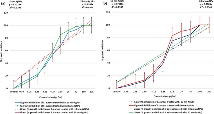
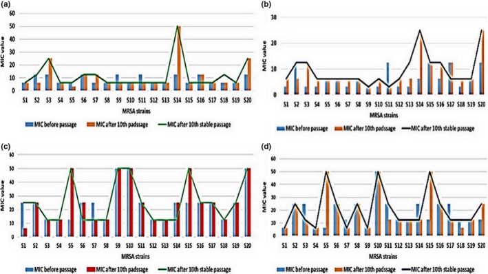
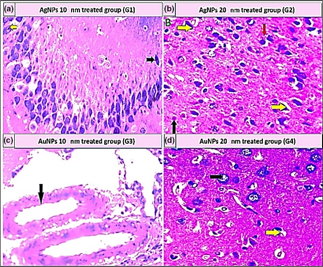
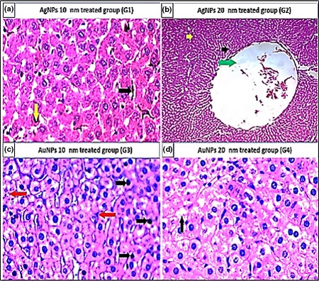
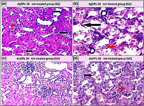
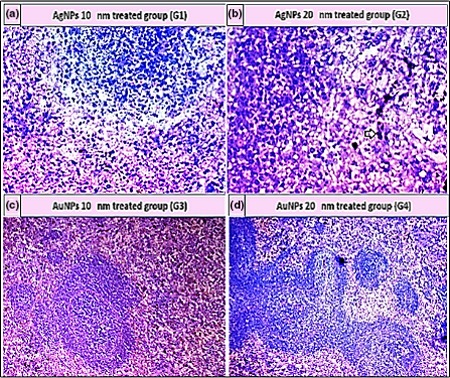
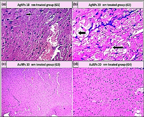
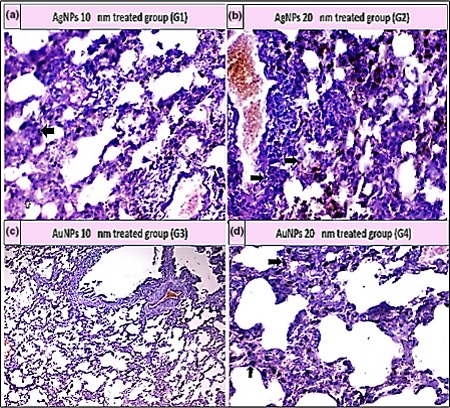
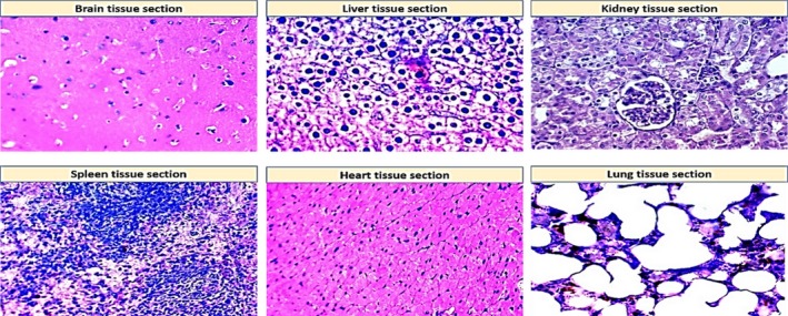
Staphylococcus aureus (S. aureus) is one of the prevalent mastitis-inducing pathogens worldwide. The resistance of S. aureus to antibiotics is a common issue for dairy farms. Recently, nanoparticles (NPs) have been used for treating antibiotic-resistant bacteria. We therefore aimed to investigate the antimicrobial effect of silver and gold NPs (AgNPs and AuNPs, respectively) and the resistance developed by S. aureus as well as the toxic effects of both NPs in rats. We used 198 S. aureus strains to determine the antibacterial effects of AgNPs and AuNPs. The microdilution method was used to establish the minimum inhibitory concentrations (MICs) of both NPs. To induce resistance, 20 S. aureus strains were passaged 10 times in broth medium with sublethal doses of NPs and an additional 10 times without NPs to examine the stability of resistance. Histopathology was performed after oral administration to the rats with the study doses of 0.25, 0.5, 1, and 2 mg/kg of NPs for 30 days. The MICs of 10-nm AgNPs, 20-nm AgNPs, 10-nm AuNPs, and 20-nm AuNPs against S. aureus were 14.70 ± 1.19 μg/ml, 9.15 ± 0.13 μg/ml, 24.06 ± 2.36 μg/ml, and 18.52 ± 1.26 μg/ml, respectively. Most strains developed strong resistance when treated with 20-nm or 10-nm AgNPs, whereas only two strains were resistant to 10-nm AuNPs and three strains to 20-nm AuNPs. No cross-resistance between NPs and various antibiotics was identified in any of the adapted S. aureus strains. Organ histopathology revealed that 0.25, 0.5, and 1 mg/kg doses of AgNPs and AuNPs were not toxic to rat tissue. In contrast, a higher dose (2 mg/kg) of NPs impaired all organs tested. This study demonstrates the antibacterial effects of NPs. S. aureus strains develop resistance less frequently against AuNPs than AgNPs, and neither AuNPs nor AgNPs was toxic to rats at low doses.

Keywords: Staphylococcus aureus; antibacterial; nanoparticles; resistance induction; toxicity.


Conflict of interest statement

The authors declare that they have no competing interests to disclose.

Figures

Figure 1
Figure 1
Antibacterial activity of AgNPs and AuNPs against Staphylococcus aureus (data represent mean ± SE of double replicates). (a) Graphical representation of % growth inhibition over concentration of 10‐ and 20‐nm AgNPs and (b) % growth inhibition over concentration of 10‐ and 20‐nm AuNPs
Figure 2
Figure 2
MIC of NPs against 20 strains of S. aureus before passage, after 10th passage, and after 10th stable passage. (a) 10‐nm AgNPs; (b) 20‐nm AgNPs; (c) 10‐nm AuNPs; and (d) 20‐nm AuNPs
Figure 3
Figure 3
Histopathological examination of brain tissue stained with H&E (400×). (a) Aggregation of 10‐nm AgNPs in the neuropil (yellow arrow); some neuronal cells appeared darkly stained (dark arrow). (b) Distorted, darkly stained neurons (yellow arrows), mild apoptosis (black arrow), and 20‐nm AgNP deposition (red arrow). (c) Large blood vessels with hyalinization of their walls in the cerebral cortex of brain and deposition of 10‐nm AuNPs (black arrow). (d) Degenerative changes in the astrocytes (black arrow) with mild apoptosis (yellow arrows)
Figure 4
Figure 4
Histopathological examination of liver tissue stained with H&E (400×). (a) Apoptosis (black arrows) and NP pigment (yellow arrow). (b) Necrosis (green arrow), apoptosis (black arrows), and NP pigment (yellow arrow). (c) Apoptosis (black arrows) and NP pigment (red arrow). (d) Hydropic degeneration (black arrows)
Figure 5
Figure 5
Histopathological examination of kidney tissue stained with H&E (400×). (a) Cloudy swelling (black arrow) and NP pigment (red arrow). (b) Marked tubular necrosis (black arrow) and NP pigment (red arrow). (c) Cloudy swelling (d) moderate tubular necrosis (black arrow) and NP pigment (red arrow)
Figure 6
Figure 6
Histopathological examination of spleen tissue stained with H&E (400×). (a) NPs deposition. (b) Marked pigment deposition (black arrow). (c) Atrophied follicles, hemorrhage, and pigmentation. (d) Marked pigment deposition (black arrow)
Figure 7
Figure 7
Histopathological examination of heart tissue stained with H&E (400×). (a) Apoptosis (black arrow) and blood vessel congestion (yellow arrow). (b) Marked hydropic degeneration (black arrow) and apoptosis. (c) Cloudy swelling and blood vessel congestion. (d) Hydropic degeneration and apoptosis
Figure 8
Figure 8
Histopathological examination of lung tissue stained with H&E (400×). (a) Moderate thickened interstitial septa and pigment deposition (black arrow); (b) Thickening of interstitial septa with marked pigment deposition (black arrow); (c) Mild thickened interstitial septa with mild pigmentation; and (d) Moderate thickened interstitial septa and pigment deposition (black arrow)
Figure 9
Figure 9
Histopathological examination of normal tissue sections for different organs (control group, G5) stained with H&E (400×)

References

    1. Ali, D. M. , Thajuddin, N. , Jeganathan, K. , & Gunasekaran, M. (2011). Plant extract mediated synthesis of silver and gold nanoparticles and its antibacterial activity against clinically isolated pathogens. Colloids and Surfaces B: Biointerfaces, 2, 360–365. - PubMed
    1. Benić, M. H. , Habrun, B. , & Kompes, G. (2012). Clinical and epidemiological aspects of cow mastitis caused by Staphylococcus aureus and its methicillin‐resistant strains. Journal of Medical Sciences, 37, 113–121.
    1. Biel, M. A. , Sievert, C. , Usacheva, M. , Teichert, M. , Wedell, E. , Loebel, N. , … Zimmermann, R. (2011). Reduction of endotracheal tube biofilms using antimicrobial photodynamic therapy. Lasers in Surgery and Medicine, 43, 586–590. 10.1002/lsm.21103 - DOI - PMC - PubMed
    1. Boschi, F. , & De Sanctis, F. (2017). Overview of the optical properties of fluorescent nanoparticles for optical imaging. European Journal of Histochemistry, 61, 245–248. - PMC - PubMed
    1. Braydich‐Stolle, L. , Hussain, S. , Schlager, J. J. , & Hofmann, M. C. (2005). In vitro cytotoxicity of nanoparticles in mammalian germline stem cells. Toxicological Sciences, 88, 412–419. 10.1093/toxsci/kfi256 - DOI - PMC - PubMed

Publication types

MeSH terms

LinkOut - more resources