

A 15-year follow up of an orthodontic treatment including a lower incisor extraction and keeping the maxillary canine-premolar transposition
- PMID: 30080123
- PMCID: PMC8111835
- DOI: 10.2319/020118-93.1
A 15-year follow up of an orthodontic treatment including a lower incisor extraction and keeping the maxillary canine-premolar transposition
Abstract
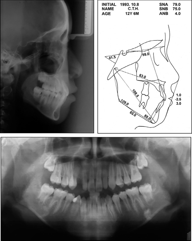
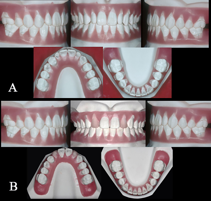
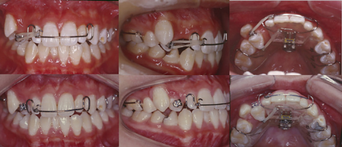
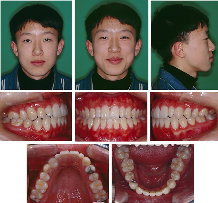
Lower incisor extraction is an effective option for treating lower anterior crowding in patients with a good facial profile, Class I molar occlusion, and narrow upper incisors. This report describes the successful treatment of an adolescent patient with lower anterior crowding and a transposed maxillary canine and premolar treated by extracting a lower incisor and keeping the transposed positions of the teeth. With the use of retainers, treatment results were stable up to the 2-year postretention visit. However, upon a 15-year postretention appointment, the fixed retainer had been removed and the removable retainer was no longer in use, which resulted in relapse of lower anterior alignment. Moreover, the transposed canine had extruded during this period, causing occlusal interference and gingival recession, as well as loss of tooth vitality, which indicates the importance of maintaining orthodontic retainers for long-term stable occlusion.
Keywords: Incisor extraction; Long term; Relapse; Retention; Transposition.
Figures


References
-
- Weintraub JA, Vig PS, Brown C, Kowalski CJ. The prevalence of orthodontic extractions. Am J Orthod Dentofacial Orthop. 1989;96:462–466. - PubMed
-
- Proffit WR. Forty-year review of extraction frequencies at a university orthodontic clinic. Angle Orthod. 1994;64:407–414. - PubMed
-
- Dacre JT. The long term effects of one lower incisor extraction. Eur J Orthod. 1985;7:136–144. - PubMed
-
- Faerovig E, Zachrisson BU. Effects of mandibular incisor extraction on anterior occlusion in adults with Class III malocclusion and reduced overbite. Am J Orthod Dentofacial Orthop. 1999;115:113–124. - PubMed
-
- Kokich VG, Shapiro PA. Lower incisor extraction in orthodontic treatment. Four clinical reports. Angle Orthod. 1984;54:139–153. - PubMed
Publication types
MeSH terms
LinkOut - more resources
Full Text Sources
Other Literature Sources
