

Utilizing 18F-FDG PET/CT Imaging and Quantitative Histology to Measure Dynamic Changes in the Glucose Metabolism in Mouse Models of Lung Cancer
- PMID: 30080208
- PMCID: PMC6126521
- DOI: 10.3791/57167
Utilizing 18F-FDG PET/CT Imaging and Quantitative Histology to Measure Dynamic Changes in the Glucose Metabolism in Mouse Models of Lung Cancer
Abstract
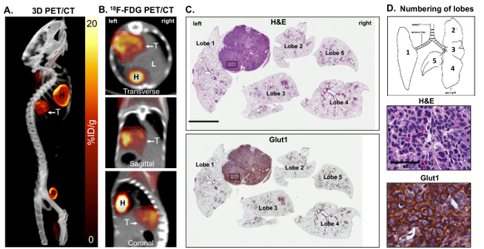
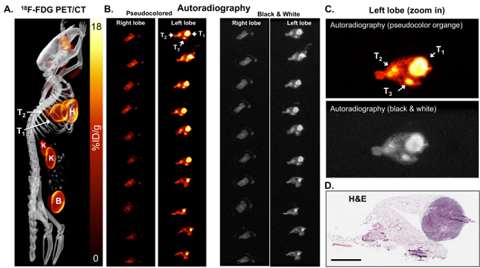
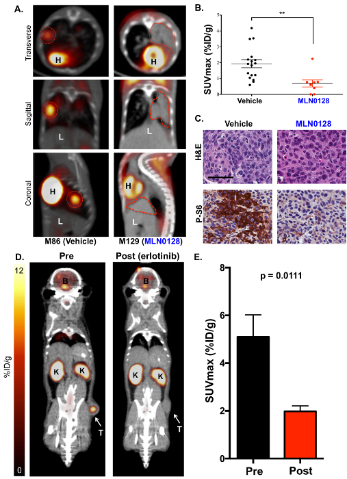
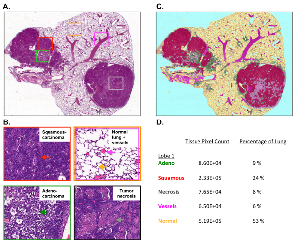
A hallmark of advanced tumors is a switch to aerobic glycolysis that is readily measured by [18F]-2-fluoro-2-deoxy-D-glucose positron emission tomography (18F-FDG PET) imaging. Co-mutations in the KRAS proto-oncogene and the LKB1 tumor suppressor gene are frequent events in lung cancer that drive hypermetabolic, glycolytic tumor growth. A critical pathway regulating the growth and metabolism of these tumors is the mechanistic target of the rapamycin (mTOR) pathway, which can be effectively targeted using selective catalytic mTOR kinase inhibitors. The mTOR inhibitor MLN0128 suppresses glycolysis in mice bearing tumors with Kras and Lkb1 co-mutations, referred to as KL mice. The therapy response in KL mice is first measured by 18F-FDG PET and computed tomography (CT) imaging before and after the delivery of MLN0128. By utilizing 18F-FDG PET/CT, researchers are able to measure dynamic changes in the glucose metabolism in genetically engineered mouse models (GEMMs) of lung cancer following a therapeutic intervention with targeted therapies. This is followed by ex vivo autoradiography and a quantitative immunohistochemical (qIHC) analysis using morphometric software. The use of qIHC enables the detection and quantification of distinct changes in the biomarker profiles following treatment as well as the characterization of distinct tumor pathologies. The coupling of PET imaging to quantitative histology is an effective strategy to identify metabolic and therapeutic responses in vivo in mouse models of disease.

References
-
- Shaw RJ, et al. The LKB1 tumor suppressor negatively regulates mTOR signaling. Cancer Cell. 2004;6(1):91–99. - PubMed
Publication types
MeSH terms
Substances
Grants and funding
LinkOut - more resources
Full Text Sources
Other Literature Sources
Medical
Miscellaneous
